. 2009 Jan;20(1):498-508.
doi: 10.1091/mbc.e08-02-0189. Epub 2008 Oct 22.

Human Discs Large is a new negative regulator of human immunodeficiency virus-1 infectivity

Affiliations

Human Discs Large is a new negative regulator of human immunodeficiency virus-1 infectivity

Fabien Perugi et al. Mol Biol Cell. 2009 Jan.

Abstract

Human immunodeficiency virus (HIV)-1 replication is positively or negatively regulated through multiple interactions with host cell proteins. We report here that human Discs Large (Dlg1), a scaffold protein recruited beneath the plasma membrane and involved in the assembly of multiprotein complexes, restricts HIV-1 infectivity. The endogenous Dlg1 and HIV-1 Gag polyprotein spontaneously interact in HIV-1-chronically infected T cells. Depleting endogenous Dlg1 in either adherent cells or T cells does not affect Gag maturation, production, or release, but it enhances the infectivity of progeny viruses five- to sixfold. Conversely, overexpression of Dlg1 reduces virus infectivity by approximately 80%. Higher virus infectivity upon Dlg1 depletion correlates with increased Env content in cells and virions, whereas the amount of virus-associated Gag or genomic RNA remains identical. Dlg1 knockdown is also associated with the redistribution and colocalization of Gag and Env toward CD63 and CD82 positive vesicle-like structures, including structures that seem to still be connected to the plasma membrane. This study identifies both a new negative regulator that targets the very late steps of the HIV-1 life cycle, and an assembly pathway that optimizes HIV-1 infectivity.


Figures
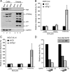
Figure 1.
Figure 1.
Dlg1 is a negative regulator of HIV-1 infectivity. (A) 293T cells were transfected with a control plasmid (Mock) or with the HIV LAI plasmid and either the control (siCtl) or anti-Dlg1 (siDlg1) siRNA, and cell lysates and supernatants were collected 72 h after transfection. Total proteins were analyzed by Western blot by using the anti-PDZ mAb for endogenous Dlg1 (top), the anti-HIV-1 human serum for Gag (middle), and the anti-GAPDH mAb for the loading control (bottom). The data are from one representative experiment out of three performed. (B and C) 293T cells (B) or MOLT-4 T cells (C) were transfected with the HIV LAI plasmid and either the control or anti-Dlg1 siRNA, and cell lysates and supernatants were collected 72 h after transfection. The amounts of intracellular (Cells) and extracellular (Virions) Gag proteins were measured using the anti-CA-p24 EIA. For infectivity, the equivalent of 1 ng of p24 was added to P4.2 cells (HeLa-HIV-LTR-lacZ) and β-galactosidase production was assessed by the CPRG method. Virus production and infectivity were normalized to control siRNA conditions (100%). The results are the mean and standard deviations of at least two independent experiments performed in duplicates. (D) 293T cells were transfected with the HIV LAI plasmid and either 0.2 or 0.5 μg of a plasmid encoding the rat version of Dlg (SAP97), and cell supernatants were harvested 24 and 48 h after transfection. The equivalent of 1 ng of p24 was added to P4.2 cells, and infectivity was measured as described above. The data represent the mean of one representative experiment out of two performed. The inset shows the production of total Dlg1 (endogenous + transfected) in cells seen by immunoblot.
Figure 2.
Figure 2.
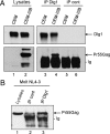
Endogenous Dlg1 interacts with Gag in HIV-1–infected T cells. (A) Cell extracts from noninfected (CEM) or HIV-1–infected (CEM-IIIB) CEM T cells were immunoprecipitated using either a rabbit serum directed to the N-terminal part of Dlg1 (IP Dlg1) or a control rabbit serum (IP Cont). Total (Lysates) or precipitated (IP) proteins were blotted using an anti-PDZ mAb for endogenous Dlg1 (top) or the anti-HIV-1 human serum for Gag (bottom). (B) Cell extracts from Molt T cells chronically infected with the NL4.3 HIV-1 strain (Molt-NL4.3) were precipitated with the control or anti-Dlg1 rabbit serum as described above, and total (Lysates) or precipitated proteins (IP) were blotted using the anti-HIV-1 human serum. Ig design the heavy chains of the antibody used for the immunoprecipitations.
Figure 3.
Figure 3.
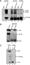
Endogenous Dlg1 does not recruit gp160, gp120, or gp41 Env proteins and is not incorporated within virions. (A) Cell extracts from noninfected (Molt) or HIV-1–infected (Molt NL4–3) Molt T cells were precipitated using control or anti-Dlg1 rabbit serum, and total (Lysates) or precipitated proteins (IP) were blotted using either the anti-gp120 110-H mAb (top) or the anti-HIV-1 human serum for Gag (bottom). (B) Cell extracts from 293T cells transfected with the HIV-1 Env expressor plasmid pMA-243 were precipitated as in A, and total (Lysates) or precipitated (IP) proteins were blotted using either the anti-PDZ mAb for endogenous Dlg1 (top) or the anti-gp41 41A mAb (bottom). (C) 293T cells were transfected with a control plasmid (Mock) or the LAI proviral plasmid and lysates from cells or ultrapurified virions were prepared 24 h after transfection. Proteins were blotted using the anti-PDZ mAb for endogenous Dlg1 (top) or the anti-p24 mAb for Gag (bottom). Ig in A and B design the heavy chains of the antibody used for the immunoprecipitations.
Figure 4.
Figure 4.
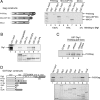
HIV-1 Gag and Dlg1 directly interact in vitro. (A) Left, Gag constructs used in the experiments. Similar amounts (see 10% input) of in vitro translated radiolabeled HIV-1 full-length Pr55Gag or truncated MACASP1NC and MACA proteins were incubated with constant amounts of immobilized GST or GST-Dlg1 proteins (right). Bound Gag proteins were resolved by SDS-PAGE and quantified by autoradiography using a Storm-850 PhosphorImager. Ten percent of the Gag input was also loaded into the gel (10% input). The percentage of binding to GST-Dlg1 normalized to the binding of full-length Gag (100%) are indicated under the autoradiography. (B) Top, result of the pull-down experiment. Bottom, amount of each GST-Gag protein used in the reaction. Similar amounts of in vitro translated radiolabeled Dlg1 were incubated with constant amount of immobilized GST alone or GST fused to Gag or Gag isolated domains as indicated. Bound Dlg1 proteins were resolved by SDS-PAGE and visualized by autoradiography using a Storm-850 PhosphorImager. Ten percent of the Dlg1 input was also loaded into the gel (10% input). (C) In vitro-translated Gag proteins were preincubated with RNase, RNase/DNase mix, or water in binding buffer before addition of GST-Dlg1. Pulled down Gag proteins and proteins from a control translation reaction (Empty) were blotted using the anti-p24 antibody. (D) Left, GST-Dlg1 constructs used in the experiments. Top, result of the pull-down experiment and the right bottom panel shows the amount of each GST-Dlg1 protein used in the reaction. Similar amounts of in vitro translated radiolabeled HIV-1 Pr55Gag were incubated with comparable amounts of immobilized GST, GST-Dlg1 or GST fused to isolated domains of Dlg1, as indicated. Bound Gag proteins were resolved by SDS-PAGE and quantified by autoradiography using a Storm-850 PhosphorImager (top left). Ten percent of the Gag input was also loaded into the gel (10% input).
Figure 5.
Figure 5.
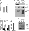
Dlg1 knockdown has not effect on gRNA packaging but increases the amount of Env in cells and virus. (A) Virus-associated RNAs prepared form equal amounts of purified particles were reverse transcribed, and the corresponding cDNA molecules were amplified using HIV-1–specific primers. The results are normalized to the amount of gRNA found in control conditions and are the means and standard deviations of two independent experiments. (B) Virus particles produced from 293T cells transfected with a control plasmid (Mock) or with the LAI plasmid and either control (siCtl) or anti-Dlg1 (siDlg1) siRNA were purified from cell supernatants by centrifugation on a 25% sucrose cushion. Total cell proteins (Cells) and virus-associated proteins (Virions) were blotted using the anti-PDZ mAb for endogenous Dlg1 (top), the 110-H mAb for Env (middle), and the anti-HIV-1 human serum for Gag (bottom). The results are from one representative experiment out of four performed. (C) Quantification of the Gag and Env protein levels of experiments presented in B. Relative virus infectivity measured in the same experiments is presented in the right histogram. The results are normalized to siRNA control conditions (100%) and are the means and standard deviations of four independent experiments. (D) 293T cells were transfected with a control plasmid (Mock) or with the HIV-1 Env expressor pMA-243 and either the control (siCtl) or anti-Dlg1 (siDlg1) siRNA. Total cell proteins were blotted with the anti-PDZ mAb for Dlg1, the 110-H mAb for gp160 and gp120, the 41A mAb for gp41, and the anti-GAPDH mAb for the loading control, as indicated.
Figure 6.
Figure 6.
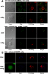
Dlg1 knockdown in T cells modulates the distribution of Gag. (A) MOLT-4 or Jurkat T cells were transfected with the pLAI plasmid and with either the control (siCtl) or anti-Dlg (siDlg1) siRNA. Seventy-two hours after transfection, cells were costained for Dlg1 (green) and Gag (red). Colocalization of green and red signals occurs as white pixels. Images are single slices from median section of cells recorded in confocal microscopy using a 63× objective. (B) CEM T cells (Dlg+/GFP−) and CEM-shDlg (Dlg−/GFP+) were infected with HIV-1 LAI and cultivated for 7 d. Cells were then costained for Dlg1 (blue) and Gag (red). Colocalization of blue and red signals occurs as mauve pixels. Images are single slices from median section of cells recorded in a confocal microscope using a 63× objective.
Figure 7.
Figure 7.
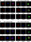
Dlg1 knockdown redistributes Gag and Env in CD63- and CD82-positive dispersed compartments. (A) Parental CEM (Dlg+/GFP−) or CEM-shDlg (Dlg−/GFP+) T cells were infected with HIV-1 LAI and cultivated for 7 d. The cells were fixed, permeabilized, and costained for Gag (red) and either CD63, CD82, or LAMP2 (blue). (B) In parallel experiments, cells were costained for Env (red) and either CD63, CD82, or LAMP2 (blue). For CEM-shDlg cells, GFP expression is also showed in the merge. Colocalization of blue and red signals occurs as mauve pixels. Images are single slices from median section of cells recorded in a confocal microscope using a 63× objective.
Figure 8.
Figure 8.
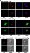
Env colocalizes with Gag in Dlg1-depleted T cells and accumulates in vesicle-like structures at the plasma membrane. (A) Parental CEM (Dlg+/GFP−) or CEM-shDlg (Dlg−/GFP+) T cells were infected with HIV-1 LAI and cultivated for 7 d. Cells were then fixed, permeabilized, and costained for Gag (red) and Env (blue). Colocalization of red and blue signals occurs as mauve pixels. (B) In parallel experiments, CEM and CEM-shDlg T cells were fixed but not permeabilized, and cell surface staining of Env was performed (red). Images are single slices from median section of cells recorded in a confocal microscope using a 63× objective.

Similar articles

Cited by

References

    1. Batonick M., Favre M., Boge M., Spearman P., Honing S., Thali M. Interaction of HIV-1 Gag with the clathrin-associated adaptor AP-2. Virology. 2005;342:190–200. - PubMed
    1. Berthoux L., Pechoux C., Ottmann M., Morel G., Darlix J. L. Mutations in the N-terminal domain of human immunodeficiency virus type 1 nucleocapsid protein affect virion core structure and proviral DNA synthesis. J. Virol. 1997;71:6973–6981. - PMC - PubMed
    1. Blot V., Delamarre L., Perugi F., Pham D., Benichou S., Benarous R., Hanada T., Chishti A. H., Dokhelar M. C., Pique C. Human Dlg protein binds to the envelope glycoproteins of human T-cell leukemia virus type 1 and regulates envelope mediated cell-cell fusion in T lymphocytes. J. Cell Sci. 2004;117:3983–3993. - PubMed
    1. Booth A. M., Fang Y., Fallon J. K., Yang J. M., Hildreth J. E., Gould S. J. Exosomes and HIV Gag bud from endosome-like domains of the T cell plasma membrane. J. Cell Biol. 2006;172:923–935. - PMC - PubMed
    1. Camus G., Segura-Morales C., Molle D., Lopez-Verges S., Begon-Pescia C., Cazevieille C., Schu P., Bertrand E., Berlioz-Torrent C., Basyuk E. The clathrin adaptor complex AP-1 binds HIV-1 and MLV Gag and facilitates their budding. Mol. Biol. Cell. 2007;18:3193–3203. - PMC - PubMed

Publication types

MeSH terms

LinkOut - more resources