. 2009 Jan;20(1):153-63.
doi: 10.1091/mbc.e08-02-0157. Epub 2008 Oct 22.

An internal domain of Exo70p is required for actin-independent localization and mediates assembly of specific exocyst components

Affiliations

An internal domain of Exo70p is required for actin-independent localization and mediates assembly of specific exocyst components

Alex H Hutagalung et al. Mol Biol Cell. 2009 Jan.

Abstract

The exocyst consists of eight rod-shaped subunits that align in a side-by-side manner to tether secretory vesicles to the plasma membrane in preparation for fusion. Two subunits, Sec3p and Exo70p, localize to exocytic sites by an actin-independent pathway, whereas the other six ride on vesicles along actin cables. Here, we demonstrate that three of the four domains of Exo70p are essential for growth. The remaining domain, domain C, is not essential but when deleted, it leads to synthetic lethality with many secretory mutations, defects in exocyst assembly of exocyst components Sec5p and Sec6p, and loss of actin-independent localization. This is analogous to a deletion of the amino-terminal domain of Sec3p, which prevents an interaction with Cdc42p or Rho1p and blocks its actin-independent localization. The two mutations are synthetically lethal, even in the presence of high copy number suppressors that can bypass complete deletions of either single gene. Although domain C binds Rho3p, loss of the Exo70p-Rho3p interaction does not account for the synthetic lethal interactions or the exocyst assembly defects. The results suggest that either Exo70p or Sec3p must associate with the plasma membrane for the exocyst to function as a vesicle tether.


Figures
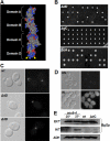
Figure 1.
Figure 1.
The N and C termini of Exo70p are essential. (A) A space-filled model of the structure of amino acid residues 62-623 of S. cerevisiae Exo70p. The domains are labeled A through D from the N terminus to the C terminus. Residues marked in blue or red indicate a positive or negative charge, respectively. The arrows point to the cluster of positively charged residues in the C terminus of Exo70p. The image was rendered using Cn3D 4.1 (NCBI) and the PDB file 2B1E (Dong et al., 2005). (B) Tetrad dissections of diploid yeast heterozygous for a deletion of the C terminus or domain D (amino acids 537-623, ΔdD), domain C (amino acids 346-515, ΔdC), and the N-terminal domain A (amino acids 1-153, ΔdA). The two viable:two inviable arrangement of spores demonstrate that the N and C termini of Exo70p are essential, whereas domain C is not essential. (C) Fluorescence microscopy of Exo70-GFP (wt) and exo70ΔdD-GFP (ΔdD) in diploid cells. Left, bright field images. Right, fluorescence images. (D) Fluorescence microscopy of GFP-exo70ΔdA in diploid cells before (UN) and after 4 h of galactose induction (IN). (E) Western blot analysis of internal and external pools of Bgl2p in wild-type versus mutant cells. To control for protein loading, Western blots against alcohol dehydrogenase (ADH) were performed.
Figure 2.
Figure 2.
Electron microscopy of wt (A) and exo70ΔdC (B) cells. Bars, 1 μm.
Figure 3.
Figure 3.
Localization of Exo70ΔdC-GFP relies on the actin cytoskeleton. (A) Exo70ΔdC-GFP localizes to sites of secretion in cells treated with DMSO but shows a diffuse localization pattern in cells treated with 100 μM LatA dissolved in DMSO. Exo70-GFP has a polarized localization in cells treated with DMSO or LatA. (B) Quantitation of Exo70ΔdC-GFP and Exo70-GFP localization from at least 100 cells for each condition. The error bars represent ± SD.
Figure 4.
Figure 4.
Point mutations in the region between residues 346 and 515 of Exo70p disrupt its interaction with Rho3p. (A) Amino acid sequence of Exo70p between residues 346 and 515. Residues in gray and underlined in gray have been mutated to alanine. The numbers below the mutated residues indicate each mutation that was created. Some of the mutations changed two adjacent charged residues (mutants 1, 2, 3, 7, 10, 13, and 15) or two charged residues separated by an uncharged residue (mutants 11 and 12) to alanine simultaneously. The residues underlined in black, K354, K355 (mutant 3) and E370 (mutant 4), respectively, disrupt the interaction of Exo70p with Rho3p when changed to alanine as tested in an in vitro binding assay shown in B. (B) In vitro binding of Exo70(K354A, K355A)p, Exo70(E370A)p, and Exo70(E499A)p to either GTPγS- or GDP-bound GST-Rho3p. (C) Structure of domain C depicting residues that disrupt the binding of Rho3p (highlighted in yellow). Those labeled with white font were identified in this study, whereas those labeled with orange font were previously identified (He et al., 2007). The structure of domain C was rendered using Cn3D 4.1 (NCBI) and PDB file 2PFV (Moore et al., 2007). (D), dissection of EXO70/exo70ΔdC; SEC3/sec3ΔN; [2μ SEC1] diploid cells. exo70ΔdC is marked with circles, sec3ΔN is marked with squares, and 2μ SEC1 is marked with diamonds. Circles or squares with dashed lines indicate comigration with the 2μ SEC1 plasmid. Eleven double mutants are predicted of which six should cosegregate with the 2μ SEC1 plasmid (based on the frequency of 15 of the 29 viable spores being SEC1+). None of the predicted double mutants are viable.
Figure 5.
Figure 5.
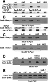
Assembly of several exocyst subunits is reduced in a exo70ΔdC background. In all of the panels, the negative control lane (NEG) is a pull-down from a strain containing either 3xHA-tagged Sec5p or 13xmyc-tagged Sec6p but no TAP-tagged exocyst component. (A) Western blots to detect Sec5p (3xHA tagged) being pulled down by either Sec8-TAP or Sec10-TAP from lysates of wt or exo70ΔdC cells. Input represents 1% of total protein. (B) Western blots detecting Sec6p (13xmyc tagged) being pulled down by Sec15-TAP from lysates of wt or exo70ΔdC cells. Input represents 1% of total protein probed with antibodies to either Sec6 (anti-myc antibodies) or Sec15 (anti-rabbit IgG antibodies). (C) Western blots detecting Sec6p (13xmyc tagged) being pulled down by either Sec8-TAP or Exo84-TAP from lysates of wt or exo70ΔdC cells. (D) Western blots detecting Sec5p (3xHA tagged) being pulled down by Sec10-TAP from lysates of wt, exo70ΔdC, exo70(K354A,K355A), or exo70(E370A) cells. (E) Western blots detecting Sec5p (3xHA tagged) pulled down with Sec8-TAP from lysates of wt, exo70ΔdC, or rho3-1 cells. In rho3-1 cells, they were grown at either 25 or 37°C for 2 h before being harvested for analysis.
Figure 6.
Figure 6.
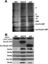
TAP tag pull-downs of the exocyst by using either Exo70-TAP or Exo70ΔdC-TAP show reduced levels of Sec6p in the assembled complex. The negative control lane (NEG) is a pull-down from a strain containing 3xHA-tagged Sec5p but no Exo70-TAP or Exo70ΔdC-TAP. (A) Silver-stained gel of the isolated exocyst complex from either wt (EXO70) cells or exo70ΔdC (ΔdC) cells. Exo70-TAP or Exo70ΔdC-TAP was pulled down, and the resulting complexes treated with TEV protease to release the exocyst complex from the IgG beads. The released exocyst complexes were then isolated on calmodulin beads and separated by SDS-polyacrylamide gel electrophoresis for silver staining. Exo70-CBP and exo70 ΔdC-CBP indicate Exo70p and Exo70 ΔdCp, respectively, with the calmodulin binding peptide after TEV cleavage. (B) Western blots detecting different exocyst components in exocyst complexes isolated from Exo70-TAP and Exo70ΔdC-TAP pull-downs. The input lanes represent 1% of total protein, and the blot was probed with anti-HA antibodies to 3xHA-tagged Sec5p.
Figure 7.
Figure 7.
Domain C of Exo70p and the N terminus of Sec3p are required for proper localization of the exocyst. The localization of Sec5-GFP is shown in sec3ΔN;exo70ΔdC;[MET17p-EXO70, CEN] (EXO70 under the inducible MET17 promoter) cells repressed for 24 h in 1 mM methionine (C and D) or subsequently induced in 0 mM methionine for 4 h (E and F). As a control, the localization of Sec5-GFP is shown in wt cells grown in 1 mM methionine (A and B), the same conditions that repress expression of EXO70 in the above-mentioned strain. (G) Quantitation of Sec5-GFP localization in sec3ΔN;exo70ΔdC;[MET17p-EXO70, CEN] with or without 1 mM methionine or wt cells treated with 1 mM methionine. At least 100 cells of each condition were counted. The error bars represent ± SD.
Figure 8.
Figure 8.
Model of the interactions between exocyst subunits mediated by Exo70p. Without residues 346-515 (domain C), the interactions between Sec5p and either Sec8p or Sec10p and Sec6p with Sec15p are reduced (indicated by dashed lines). Domain C does not seem to affect the interaction of Exo70p with the remaining exocyst components (indicated by solid lines). The exo70ΔdC mutant may reveal the function of Exo70p in bridging different exocyst subcomplexes.

Similar articles

Cited by

References

    1. Abramoff M. D., Magelhaes P. J., Ram S. J. Image processing with ImageJ. Biophotonics Int. 2004;11:36–42.
    1. Adamo J., Rossi G., Brennwald P. The Rho GTPase Rho3 has a direct role in exocytosis that is distinct from its role in actin polarity. Mol. Biol. Cell. 1999;10:4121–4133. - PMC - PubMed
    1. Ayscough K., Stryker J., Pokala N., Sanders M., Crews P., Drubin D. High rates of actin filament turnover in budding yeast and roles for actin in establishment and maintenance of cell polarity revealed using the actin inhibitor latrunculin-A. J. Cell Biol. 1997;137:399–416. - PMC - PubMed
    1. Boyd C., Hughes T., Pypaert M., Novick P. Vesicles carry most exocyst subunits to exocytic sites marked by the remaining two subunits, Sec3p and Exo70p. J. Cell Biol. 2004;167:889–901. - PMC - PubMed
    1. Dong G., Hutagalung A., Fu C., Novick P., Reinisch K. The structures of exocyst subunit Exo70p and the Exo84p C-terminal domains reveal a common motif. Nat. Struct. Mol. Biol. 2005;12:1094–1100. - PubMed

MeSH terms