

Dopamine type-1 receptor binding in major depressive disorder assessed using positron emission tomography and [11C]NNC-112
- PMID: 18946469
- PMCID: PMC2656589
- DOI: 10.1038/npp.2008.194
Dopamine type-1 receptor binding in major depressive disorder assessed using positron emission tomography and [11C]NNC-112
Abstract
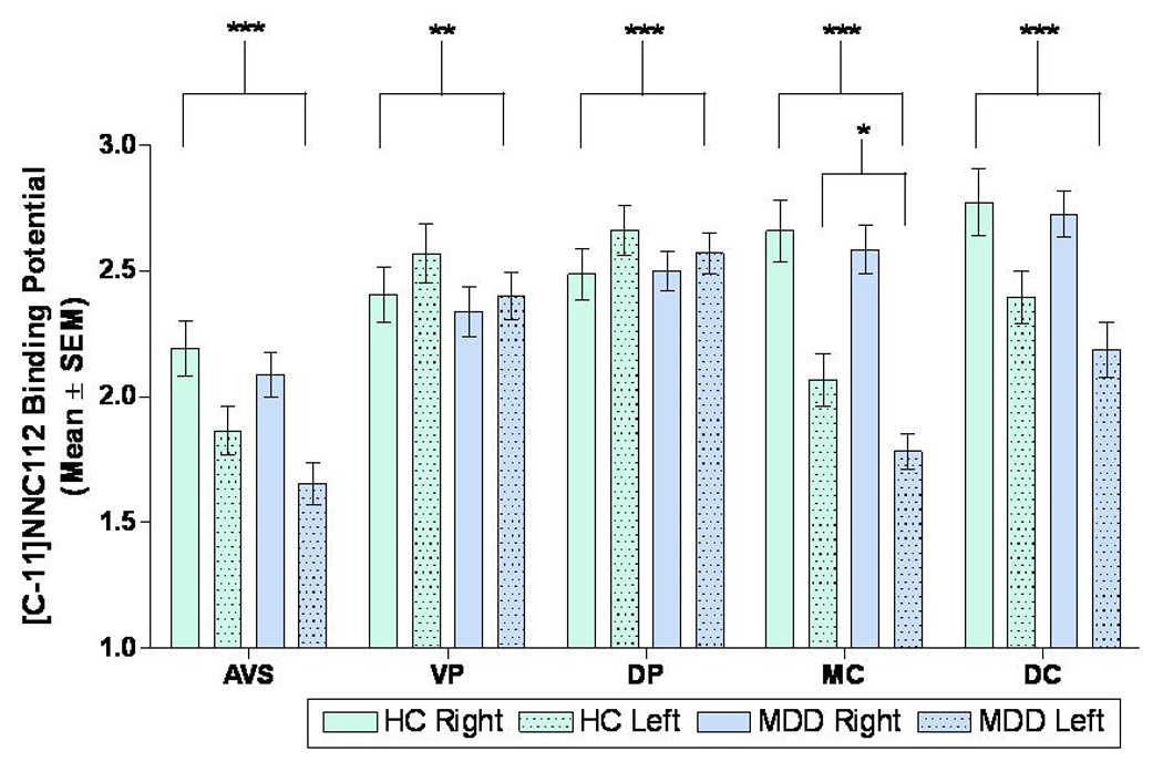
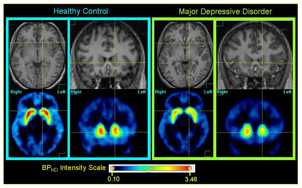
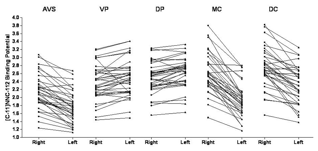
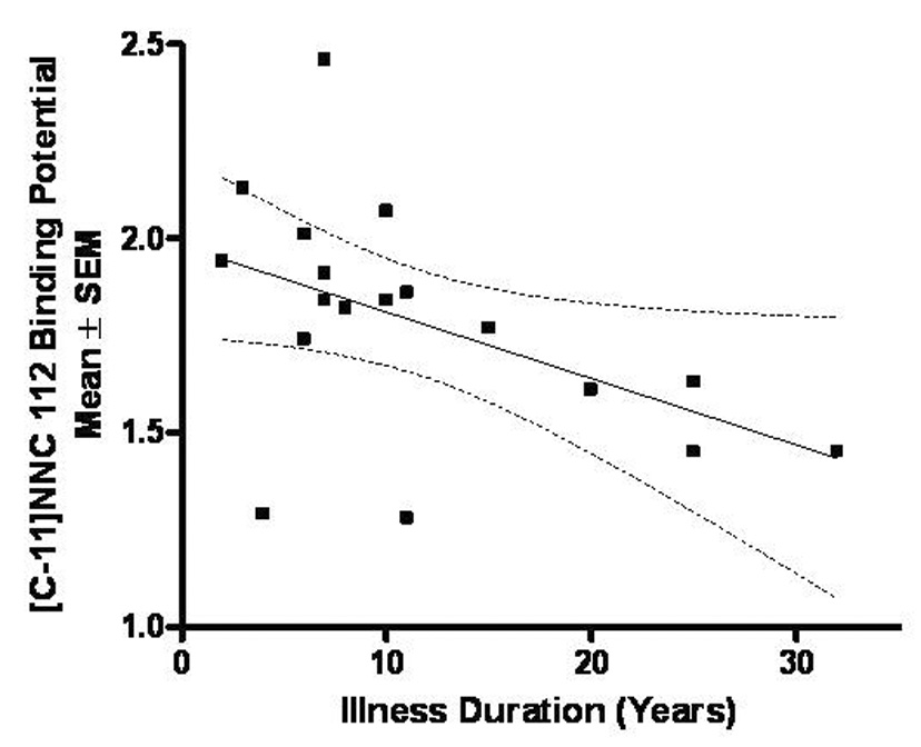
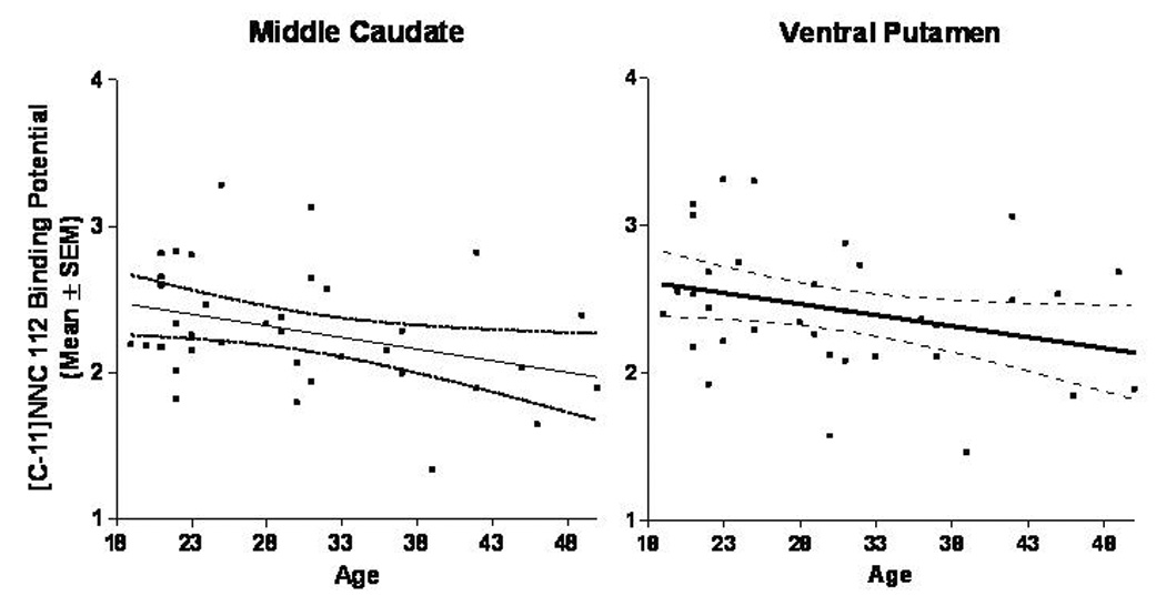
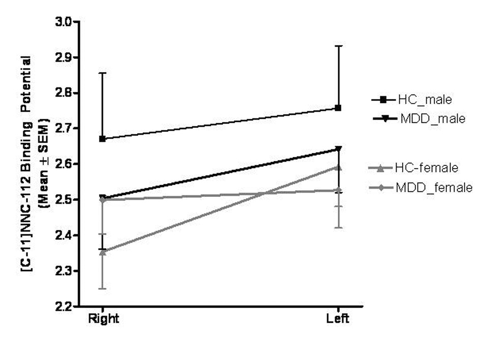
The dopamine type-1 receptor has been implicated in major depressive disorder (MDD) by clinical and preclinical evidence from neuroimaging, post mortem, and behavioral studies. To date, however, selective in vivo assessment of D(1) receptors has been limited to the striatum in MDD samples manifesting anger attacks. We employed the PET radioligand, [(11)C]NNC-112, to selectively assess D(1) receptor binding in extrastriatal and striatal regions in a more generalized sample of MDD subjects. The [(11)C]NNC-112 nondisplaceable binding potential (BP(ND)) was assessed using PET in 18 unmedicated, currently depressed subjects with MDD and 19 healthy controls, and compared between groups using MRI-based region-of-interest analysis. The mean D(1) receptor BP(ND) was reduced (14%) in the left middle caudate of the MDD group relative to control group (p<0.05). Among the MDD subjects D(1) receptor BP(ND) in this region correlated negatively with illness duration (r=-0.53; p=0.02), and the left-to-right BP(ND) ratio correlated inversely with anhedonia ratings (r=-0.65, p=0.0040). The D(1) receptor BP(ND) was strongly lateralized in striatal regions (p<0.002 for main effects of hemisphere in accumbens area, putamen, and caudate). In post hoc analyses, a group-by-hemisphere-by-gender interaction was detected in the dorsal putamen, which was accounted for by a loss of the normal asymmetry in depressed women (F=7.33, p=0.01). These data extended a previous finding of decreased striatal D(1) receptor binding in an MDD sample manifesting anger attacks to a sample selected more generally according to MDD criteria. Our data also more specifically localized this abnormality in MDD to the left middle caudate, which is the target of afferent neural projections from the orbitofrontal and anterior cingulate cortices where neuropathological changes have been reported in MDD. Finally, D(1) receptor binding was asymmetrical across hemispheres in healthy humans, compatible with evidence that dopaminergic function in the striatum is lateralized during reward processing, voluntary movement, and self-stimulation behavior.
Conflict of interest statement
Figures
Similar articles
-
Decreased striatal D1 binding as measured using PET and [11C]SCH 23,390 in patients with major depression with anger attacks.Depress Anxiety. 2006;23(3):175-7. doi: 10.1002/da.20168. Depress Anxiety. 2006. PMID: 16528700
-
Dopamine Release in Antidepressant-Naive Major Depressive Disorder: A Multimodal [11C]-(+)-PHNO Positron Emission Tomography and Functional Magnetic Resonance Imaging Study.Biol Psychiatry. 2018 Oct 15;84(8):563-573. doi: 10.1016/j.biopsych.2018.05.014. Epub 2018 May 25. Biol Psychiatry. 2018. PMID: 30041971 Free PMC article.
-
Elevated serotonin transporter binding in major depressive disorder assessed using positron emission tomography and [11C]DASB; comparison with bipolar disorder.Biol Psychiatry. 2007 Oct 15;62(8):870-7. doi: 10.1016/j.biopsych.2007.03.016. Epub 2007 Aug 2. Biol Psychiatry. 2007. PMID: 17678634
-
Pre- and post-synaptic dopamine imaging and its relation with frontostriatal cognitive function in Parkinson disease: PET studies with [11C]NNC 112 and [18F]FDOPA.Psychiatry Res. 2008 Jul 15;163(2):171-82. doi: 10.1016/j.pscychresns.2007.11.003. Epub 2008 May 27. Psychiatry Res. 2008. PMID: 18504119
-
Cerebral metabolism in major depressive disorder: a voxel-based meta-analysis of positron emission tomography studies.BMC Psychiatry. 2014 Nov 19;14:321. doi: 10.1186/s12888-014-0321-9. BMC Psychiatry. 2014. PMID: 25407081 Free PMC article. Review.
Cited by
-
Gray matter volume alterations in subjects with overweight and obesity: Evidence from a voxel-based meta-analysis.Front Psychiatry. 2022 Sep 26;13:955741. doi: 10.3389/fpsyt.2022.955741. eCollection 2022. Front Psychiatry. 2022. PMID: 36226110 Free PMC article.
-
Neurocircuitry of mood disorders.Neuropsychopharmacology. 2010 Jan;35(1):192-216. doi: 10.1038/npp.2009.104. Neuropsychopharmacology. 2010. PMID: 19693001 Free PMC article. Review.
-
Dopamine receptor alterations in female rats with diet-induced decreased brain docosahexaenoic acid (DHA): interactions with reproductive status.Nutr Neurosci. 2010 Aug;13(4):161-9. doi: 10.1179/147683010X12611460764282. Nutr Neurosci. 2010. PMID: 20670471 Free PMC article.
-
The molecular mechanism of chronic stress affecting the occurrence and development of breast cancer and potential drug therapy.Transl Oncol. 2022 Jan;15(1):101281. doi: 10.1016/j.tranon.2021.101281. Epub 2021 Dec 4. Transl Oncol. 2022. PMID: 34875482 Free PMC article. Review.
-
Recognizing and Treating Major Depression in Fibromyalgia: A Narrative Primer for the Non-Psychiatrist.J Prim Care Community Health. 2024 Jan-Dec;15:21501319241281221. doi: 10.1177/21501319241281221. J Prim Care Community Health. 2024. PMID: 39279389 Free PMC article. Review.
References
-
- Abi-Dargham A, Martinez D, Mawlawi O, Simpson N, Hwang DR, Slifstein M, Anjilvel S, Pidcock J, Guo NN, Lombardo I, Mann JJ, Van Heertum R, Foged C, Halldin C, Laruelle M. Measurement of striatal and extrastriatal dopamine D1 receptor binding potential with [11C]NNC 112 in humans: validation and reproducibility. J Cereb Blood Flow Metab. 2000;20(2):225–243. - PubMed
-
- Agren H, Reibring L. PET studies of presynaptic monoamine metabolism in depressed patients and healthy volunteers. Pharmacopsychiatry. 1994;27(1):2–6. - PubMed
-
- Andersen PH, Gronvald FC, Hohlweg R, Hansen LB, Guddal E, Braestrup C, Nielsen EB. NNC-112, NNC-687 and NNC-756, new selective and highly potent dopamine D1 receptor antagonists. Eur J Pharmacol. 1992;219(1):45–52. - PubMed
-
- APA. Diagnostic and Statistical Manual of Mental Disorders (DSM-IV) Washington, D.C.: APA Press; 1994.
-
- Argyelan M, Szabo Z, Kanyo B, Tanacs A, Kovacs Z, Janka Z, Pavics L. Dopamine transporter availability in medication free and in bupropion treated depression: a 99mTc-TRODAT-1 SPECT study. J Affect Disord. 2005;89(1–3):115–123. - PubMed
Publication types
MeSH terms
Substances
Grants and funding
LinkOut - more resources
Full Text Sources
