. 2008 Nov 19;27(22):3069-80.
doi: 10.1038/emboj.2008.223. Epub 2008 Oct 23.

Thrombospondin-1 binds to ApoER2 and VLDL receptor and functions in postnatal neuronal migration

Affiliations

Thrombospondin-1 binds to ApoER2 and VLDL receptor and functions in postnatal neuronal migration

Sophia M Blake et al. EMBO J. .

Erratum in

  • EMBO J. 2008 Dec 17;27(24):3332

Abstract

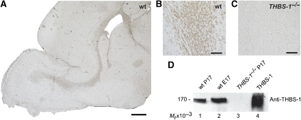
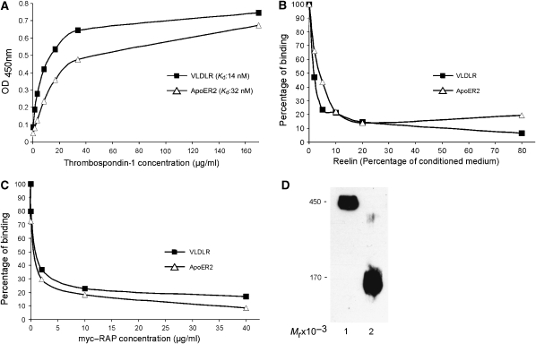
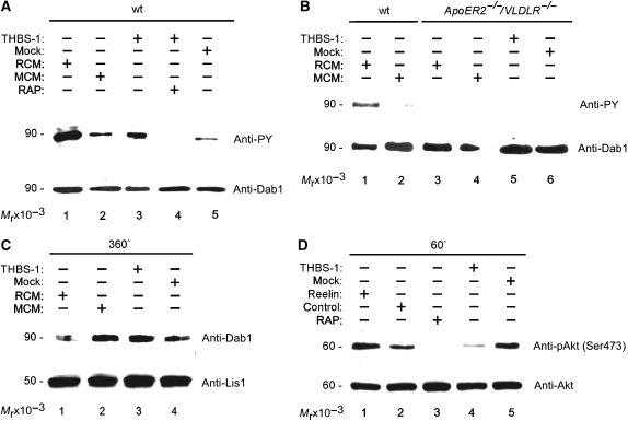
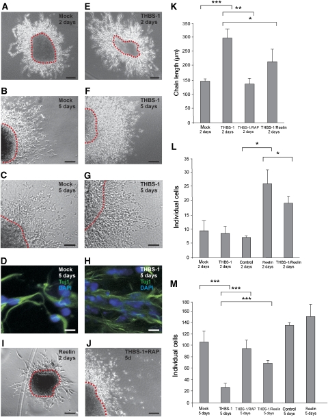
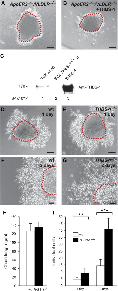
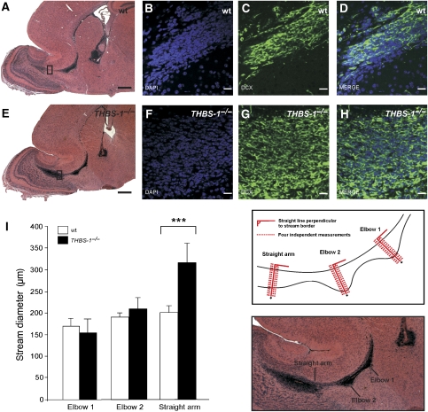
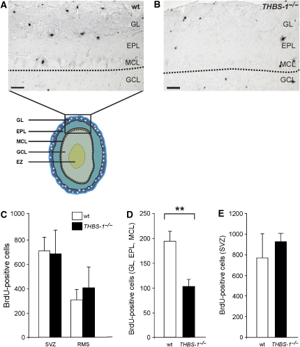
Apolipoprotein E receptor 2 (ApoER2), very low-density lipoprotein receptor (VLDLR), and Dab1 are the main components of the Reelin signalling cascade. Reelin is the sole ligand defined so far in signalling through this pathway. Postnatal migration of neuronal precursors from the subventricular zone (SVZ) to the olfactory bulb (OB), however, depends on ApoER2 and Dab1, but functions independently of Reelin. Here, we show that thrombospondin-1 (THBS-1) is a novel physiological ligand for ApoER2 and VLDLR. THBS-1 is present in the SVZ and along the entire rostral migratory stream (RMS). It binds to ApoER2 and VLDLR and induces phosphorylation of Dab1. In contrast to Reelin, it does not induce Dab1 degradation or Akt phosphorylation, but stabilizes neuronal precursor chains derived from subventricular explants. Lack of THBS-1 results in anatomical abnormalities of the RMS and leads to a reduction of postnatal neuronal precursors entering the OB.


Figures

Figure 1
Figure 1
Thrombospondin-1 is expressed in the RMS and the SVZ of postnatal mice. Sagittal sections (6 μm) of the forebrains from (A, B) wt and (C) THBS-1−/− mice were immunostained with a monoclonal antibody against THBS-1. (D) Total protein extracts from wt P17 (lane 1), wt E17 (lane 2), and THBS-1−/− P17 brains (lane 3) were prepared. The samples (20 μg of total protein lysate per lane) and purified THBS-1 (0.5 μg, lane 4) were subjected to 6% SDS–PAGE under reducing conditions, and subsequent western blot analysis was performed using a monoclonal antibody against THBS-1 and an appropriate HRP-conjugated secondary antibody. Scale bars: (A) 500 μm; (B, C) 50 μm.
Figure 2
Figure 2
Thrombospondin-1 binds to ApoER2 and VLDLR. (A) Microtitre plates were coated with recombinant ligand-binding domains of ApoER2 and VLDLR (ApoER2Δ4-6-MBP/His and VLDLR1-8-MBP/His, respectively) and incubated with the indicated amounts of THBS-1. (B, C) Microtitre plates were coated with ApoER2Δ4-6-MBP/His or VLDLR1-8-MBP/His, and incubated with THBS-1 (15 μg/ml for ApoER2 and 6.5 μg/ml for VLDLR) in the presence of increasing amounts of Reelin-conditioned medium (B) or myc-tagged RAP (myc–RAP) (C). Bound THBS-1 was detected with a monoclonal anti-THBS-1 antibody and an appropriate HRP-conjugated secondary antibody. OD450, optical density at 450 nm. Kd, dissociation constant. In each case (A–C), the results of a representative experiment are shown. Kd values have been calculated from three independent experiments. (D) Western blot analysis of purified THBS-1. THBS-1 was purified from human plasma as described in Materials and methods and subjected to 6% SDS–PAGE (0.5 μg of protein per lane) under non-reducing (lane 1) and reducing conditions (lane 2) and subsequent western blot analysis using a monoclonal antibody against THBS-1 and an appropriate HRP-conjugated secondary antibody.
Figure 3
Figure 3
Thrombospondin-1 induces Dab1 phosphorylation but not Dab1 degradation and Akt phosphorylation in primary embryonic neurons. (A) Primary mouse E16 wt neurons were incubated with Reelin-conditioned medium (RCM, lane 1), mock-conditioned medium (MCM, lane 2), purified THBS-1 (10 μg/ml, lane 3), THBS-1 in the presence of myc–RAP (lane 4), and mock medium (lane 5). Cells were processed for immunoprecipitation of Dab1, and western blotting was subsequently performed using antiphosphotyrosine (PY) or anti-Dab1 (Dab1) antibody as indicated. (B) Primary mouse E16 wt and primary E16 ApoER2−/−/VLDLR−/− neurons were incubated with RCM (lanes 1 and 3), MCM (lanes 2 and 4), purified THBS-1 (lane 5) and mock medium (lane 6). Dab1 phosphorylation was measured as indicated above. (C) Primary mouse E16 wt neurons were incubated with RCM (lane 1), MCM (lane 2), purified THBS-1 (10 μg/ml, lane 3), and mock medium (lane 4) for 6 h. Total cell extracts were prepared and western blotting was performed using anti-Dab1 and anti-Lis1 antibodies. (D) Primary mouse E16 wt neurons were incubated with Reelin (lane 1), control medium (lane 2), control medium and myc–RAP (lane 3), purified THBS-1 (10 μg/ml, lane 4), and mock medium (lane 5) for 1 h. Cell extracts were prepared and western blotting was performed using anti-phospho-Akt and anti-Akt antibodies.
Figure 4
Figure 4
Thrombospondin-1 stabilizes chains of migratory neuronal precursors from explants of the SVZ. (AD) SVZ explants were prepared from P7 wt mice and treated with mock medium, or (E–H) cultivated in the presence of 4 μg/ml purified THBS-1, (I) Reelin or (J) THBS-1 in the presence of myc–RAP for 2 days (A, E, I) or 5 days (B, C, D, F, G, H, J), respectively. (D, H) Explants were immunostained using antibodies against Tuj1 with the appropriate secondary Alexa-Fluor antibody and DAPI. Representative explants are shown. (K) Explants were analysed by measuring the length of migratory chains after 2 days in culture and the number of individual cells per field after (L) 2 days and after (M) 5 days in culture (chain length 2 days: mock: n=8, THBS-1: n=13, THBS-1/myc–RAP: n=7, THBS-1/Reelin: n=6; dissociated neurons 2 days: mock: n=5, THBS-1: n=7, control: n=2, Reelin: n=2, THBS-1/Reelin: n=4; dissociated neurons 5 days: mock: n=6, THBS-1: n=28, THBS-1/myc–RAP: n=6, THBS-1/Reelin: n=6, control: n=4, Reelin: n=4; individual explants were derived from at least four different wt mice). Plots show average+s.e.m.; ***P<0.001; **P<0.01; *P<0.05 (Student's t-test); scale bars: (A, B, E, F, I, J) 100 μm; (C, G) 50 μm; (D, H) 10 μm.
Figure 5
Figure 5
Stability of SVZ explants is dependent on THBS-1. (A) P7 ApoER2−/−/VLDLR−/− SVZ explants were treated with control medium or (B) cultivated in the presence of 4 μg/ml purified THBS-1 for 2 days. (C) Protein extracts (45 μg) from SVZ explants from wt (lane 1), THBS-1−/− (lane 2) mice, and purified THBS-1 (0.5 μg) were subjected to 6% SDS–PAGE under reducing conditions. Subsequent western blot analysis was performed using a monoclonal antibody against THBS-1 and an appropriate HRP-conjugated secondary antibody. (D, F) SVZ explants were prepared from P7 wt and (E, G) THBS-1−/− mice and kept in culture for 1 day (D, E) and 2 days (F,G). Representative explants are shown. (H) Explants were analysed by measuring the length of migratory chains after 2 days in culture and (I) the number of individual cells per field after 1 and 2 days (chain length 2 days: wt: n=5, THBS-1−/−: n=6; individual neurons 1 day: wt: n=7, THBS-1−/−: n=7; individual neurons 2 days: wt: n=17, THBS-1−/−: n=10; individual explants were derived from at least five different wt and THBS-1−/− mice). Plots show average+s.e.m.; ***P<0.001; **P<0.01; (Student's t-test); scale bars: (A, B, D, E) 100 μm; (F, G) 50 μm.
Figure 6
Figure 6
The architecture of the RMS of THBS-1−/− mice is altered. (AD) Matched sagittal sections (6 μm) of the forebrains from wt mice and (EH) THBS-1−/− mice were stained with (A, E) haematoxylin and eosin, (B, F) DAPI, or (C, G) immunostained with anti-doublecortin (DCX). (I) Measurement of the width of the RMS from wt mice (empty bars) and THBS-1−/− mice (black bars). Matched serial sections from (P17) wt (n=4) and THBS-1−/− mice (n=8) were used to measure the width of the RMS at three defined sites as described in Materials and methods. Plots show average+s.e.m., ***P<0.001 (Student's t-test); scale bars: (A, E) 500 μm; (B–D, F–H) 20 μm.
Figure 7
Figure 7
Number of newly generated neuronal precursors derived from the RMS is reduced in the OB of THBS-1−/− mice. (A) wt and (B) THBS-1−/− mice received a BrdU pulse at P12, and brains were collected at P17. Matched sagittal sections of the forebrains were stained for BrdU-positive cells. Borders between granule cell layer (GCL) and mitral cell layer (MCL) are marked by dotted lines. (C) Quantification of BrdU-positive cells in the SVZ and RMS. (D) Quantification of BrdU-positive cells in the glomerular layer (GL), ependymal layer (EPL), and MCL of wt (empty bar) and THBS-1−/− mice (black bar). (E) Quantification of BrdU-positive cells in the SVZ after a 2 h BrdU pulse. Representative sections are shown (wt mice: empty bars, THBS-1−/− mice: black bars; BrdU 5 days, SVZ and RMS: wt: n=4, THBS-1−/− n=4; BrdU 5 days, GL, EPL, and MCL: wt: n=3, THBS-1−/− n=6; BrdU 2 h, SVZ: wt: n=4, THBS-1−/−:n=3). Plots show average+s.e.m., **P<0.01 (Student's t-test); scale bars: 50 μm.

Similar articles

Cited by

References

    1. Adams JC, Tucker RP (2000) The thrombospondin type 1 repeat (TSR) superfamily: diverse proteins with related roles in neuronal development. Dev Dyn 218: 280–299 - PubMed
    1. Agah A, Kyriakides TR, Lawler J, Bornstein P (2002) The lack of thrombospondin-1 (TSP1) dictates the course of wound healing in double-TSP1/TSP2-null mice. Am J Pathol 161: 831–839 - PMC - PubMed
    1. Andrade N, Komnenovic V, Blake SM, Jossin Y, Howell B, Goffinet A, Schneider WJ, Nimpf J (2007) ApoER2/VLDL receptor and Dab1 in the rostral migratory stream function in postnatal neuronal migration independently of Reelin. Proc Natl Acad Sci USA 104: 8508–8513 - PMC - PubMed
    1. Arnaud L, Ballif BA, Forster E, Cooper JA (2003) Fyn tyrosine kinase is a critical regulator of Disabled-1 during brain development. Curr Biol 13: 9–17 - PubMed
    1. Bajari TM, Strasser V, Nimpf J, Schneider WJ (2005) LDL receptor family: isolation, production, and ligand binding analysis. Methods 36: 109–116 - PubMed

Publication types

MeSH terms