. 2008 Nov 19;27(22):3024-35.
doi: 10.1038/emboj.2008.221. Epub 2008 Oct 23.

RNA-dependent recruitment of the origin recognition complex

Affiliations

RNA-dependent recruitment of the origin recognition complex

Julie Norseen et al. EMBO J. .

Abstract

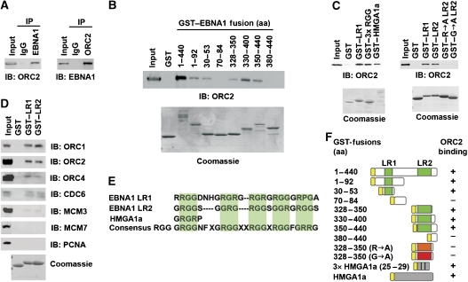
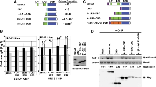
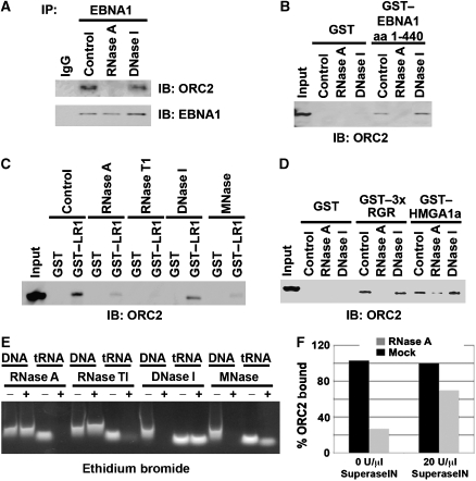
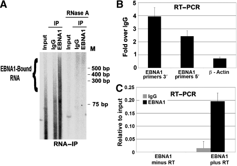
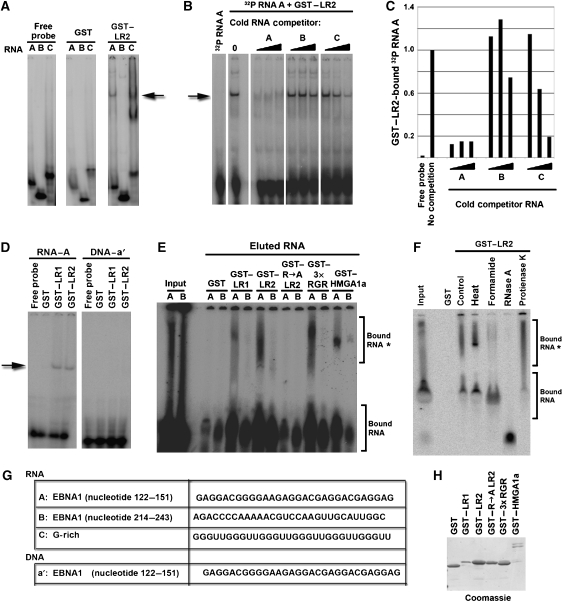
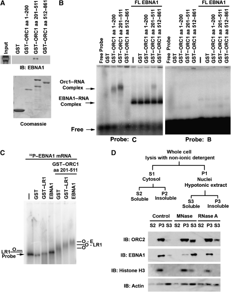
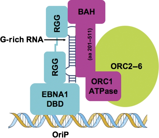
The origin recognition complex (ORC) has an important function in determining the initiation sites of DNA replication. In higher eukaryotes, ORC lacks sequence-specific DNA binding, and the mechanisms of ORC recruitment and origin determination are poorly understood. ORC is recruited with high efficiency to the Epstein-Barr virus origin of plasmid replication (OriP) through a complex mechanism involving interactions with the virus-encoded EBNA1 protein. We present evidence that ORC recruitment to OriP and DNA replication function depends on RGG-like motifs, referred to as LR1 and LR2, in the EBNA1 amino-terminal domain. Moreover, we show that LR1 and LR2 recruitment of ORC is RNA dependent. HMGA1a, which can functionally substitute for LR1 and LR2 domain, can also recruit ORC in an RNA-dependent manner. EBNA1 and HMGA1a RGG motifs bound to structured G-rich RNA, as did ORC1 peptides, which interact with EBNA1. RNase A treatment of cellular chromatin released a fraction of the total ORC, suggesting that ORC association with chromatin, and possibly cellular origins, is stabilized by RNA. We propose that structural RNA molecules mediate ORC recruitment at some cellular and viral origins, similar to OriP.


Figures

Figure 1
Figure 1
EBNA1-linking regions 1 and 2 are necessary and sufficient for ORC interaction. (A) Raji cell extracts were immunoprecipitated with anti-EBNA1antibody or IgG control (left panel) or anti-ORC2 antibody and IgG control (right panel). IPs were analysed by immunoblot with anti-ORC2 (left panel) and anti-EBNA1 (right panel). Input represents ∼5% of the total input for each IP. (B) GST–EBNA1 fusion peptides were assayed for their ability to recruit ORC2 from HeLa nuclear extracts by immunoblot with anti-ORC2. Purified GST fusion proteins were assayed by Coomassie Blue staining of SDS–PAGE gels (lower panels). EBNA1 amino-acid residues fused to GST are indicated above each lane. (C) GST fusion proteins (GST, GST–LR1 (aa 30–53), GST–3 × RGR, GST–HMGA1a, GST–LR2 (aa 328–350), GST–LR2 (R → A), or GST–LR2 (G → A) were assayed for their ability to recruit ORC2 from HeLa nuclear extracts. ORC2 recruitment was assayed by immunoblot with anti-ORC2 antibody. GST fusion protein expression and purity was assayed by Coomassie blue staining (lower panels). (D) GST–LR1, GST–LR2, or GST alone were incubated with HeLa nuclear extract and assayed by immunoblot for ORC1, ORC2, ORC4, CDC6, MCM3, MCM7, or PCNA, as indicated. Coomassie stain of GST fusion peptides is shown in the lower panel. (E) Sequence alignment of EBNA1 LR1, LR2, HGMA1a RGR, and consensus RGG domain. (F) Summary of GST fusion proteins and their ability to bind ORC2 as represented by experiments shown in panels B and C.
Figure 2
Figure 2
LR1 or LR2 confers ORC recruitment and replication at OriP. (A) Schematic of EBNA1 and fusion peptide constructs used in colony formation assay. The light blue box represents the EBNA1 nuclear localization signal (NLS). One, two, or three copies of LR1 (green boxes) were fused to EBNA1 DBD and co-transfected with plasmid-containing OriP and Puromycin-resistance gene (Puro). The number of bacterial colonies transformed with recovered plasmid OriP were quantified as an average of several experiments relative to wt EBNA1 protein. (B) Puromycin-resistant 293T cells pools generated with EBNA1 wt, 1 × LR1–DBD, 2 × LR1–DBD, or 3 × LR1–DBD were harvested for ChIP 14 days post-transfection. Enrichment of EBNA1 and ORC at OriP (black bars) or Puromycin resistance gene (grey bars) was compared with IgG controls by real-time PCR. DBD alone is not shown, as no OriP DNA was detected among the few puromycin-resistant colonies. ChIP data are presented as the log 2 of Ct values relative to IgG controls. The relative enrichment of ORC2 binding at OriP relative to the Puro gene is indicated by the numbers above each set of bars in the panel to the right. P-value of <0.05 using single ANOVA analysis are indicated by *. Expression levels for EBNA1-derived proteins are shown in the western blot to the right. (C) Schematic of fusion peptide constructs used for transient replication assay. The magenta box represents the N-terminal FLAG-epitope. (D) EBNA1 fusion peptides described in C were co-transfected with OriP-containing plasmid. DNA was harvested 72 h post-transfection and visualized with OriP-specific probe by Southern blot hybridization. Replication was measured as resistance to DpnI restriction digestion. Replication efficiency is calculated as DpnI resistance over input (BamHI) and normalized to full-length EBNA1 as quantified by ImageQuant software. Immunoblot below shows protein expression levels for FLAG-tagged EBNA1 proteins.
Figure 3
Figure 3
RNA-dependent recruitment of ORC by EBNA1 LR and HMGA1a RGR domains. (A) EBNA1 IP from Raji cells were incubated with 0.02 mg/ml RNase A, 0.2 U/ml DNase I, or buffer control. ORC2 recruitment was monitored by immunoblot (IB) with anti-ORC2 (top panel) or anti-EBNA1 (lower panel). (B) GST or GST–EBNA1 1–440 (containing LR1 and LR2) was incubated with HeLa nuclear extracts and then treated with 0.02 mg/ml RNase A, 0.2 U/ml DNase I, or buffer control. ORC2 recruitment was monitored by IB. (C) GST or GST–LR1 were incubated with HeLa nuclear extract and then treated with either 0.02 mg/ml RNase A, 2 U/ml RNase T1, 0.2 U/ml DNase I, 0.2 U/ml micrococcal nuclease (MNase I), or buffer control. ORC2 recruitment was monitored by IB with anti-ORC2. (D) GST, GST–3 × RGR, or GST–HMGA1a was incubated with HeLa nuclear extracts and then treated with 0.02 mg/ml RNase A, 0.2 U/ml DNase I, or buffer control and monitored by IB with anti-ORC2. (E) Control digestions were preformed using 50 μg of single-stranded, sonicated, heat-denatured salmon sperm DNA (ssDNA) or 100 μg of tRNA under the same conditions for RNase A, RNase TI, DNase I, and MNase I used in the IPs and GST pull-downs mentioned above. ssDNA and tRNA were separated by agarose gel electrophoresis and visualized after ethidium bromide staining. (F) RNase A-dependent loss of ORC2 binding to GST or GST–LR2 was tested by the addition of SuperaseIN (20 U/μl) in reactions containing either 0 or 90 ng/ml RNase A, as indicated. ORC2 binding was measured by western blot and quantified using ImageQuant and presented as percentage of ORC2 binding in the absence of RNase A.
Figure 4
Figure 4
EBNA1 binds its own mRNA. (A) RNA isolated from Raji cell extracts (input) or from immunoprecipitates with anti-EBNA1 or control IgG was radiolabelled with 32P-γATP and polynucleotide kinase. Labelled RNA was visualized by autoradiography of native polyacrylamide gels. RNase A-treated samples are shown on the right, as indicated. (B) RT–PCR with RNA purified from EBNA1 IPs from Raji cells. IP-derived RNA was amplified with primers specific for the EBNA1 ORF 5′ or 3′ or with primers for cellular β-actin mRNA. (C) RT–PCR with RNA purified from EBNA1 or control IgG IP using primers for EBNA1 ORF 5′-region. Reactions were performed with RT (plus) or without RT (minus) as indicated.
Figure 5
Figure 5
ORC recruiting modules bind G-rich RNA. (A) Purified GST and GST–LR2 were assayed for binding to 32P RNA oligonucleotides A, B, or C using EMSA. RNA-bound complex is indicated by the arrow. (B) Specificity of the interaction between GST–LR2 and 32P-labelled RNA probe A was assayed by addition of 3 × , 9 × , or 27 × fold molar excess of cold competitor RNAs A, B, or C to the binding reaction. The major complex formed between 32P RNA A and GST–LR2 is indicated by the arrow. (C) Graphical quantification of bound RNA A by GST–LR2 from the competition assay in panel B using ImageQuant software analysis. (D) GST, GST–LR1, and GST–LR2 were assayed for binding to 32P-labelled RNA A (left panel), or single-stranded DNA a′ of the same sequence (right panel). (E) 32P-labelled RNA A or B was eluted from GST pull-down experiments with GST, or GST fused to LR1, LR2, R → A LR2, 3 × RGR, and HMGA1a and assayed by gel electrophoresis and visualized by PhosphorImager. Input RNA is indicated in lanes 1 and 2. (F) 32P-labelled RNA A was eluted from GST pull-down with GST or GST–LR2, and then treated with heat (95°C, 3 min), formamide (80%), RNase A (0.2 mg/ml), or Protease K (0.1 mg/ml), as indicated. (G) Sequence of the RNA species A, B, and C and DNA species a′ used in these experiments. (H) Proteins used in the RNA pull-down experiment in E were separated by SDS–PAGE and visualized by Coomassie staining.
Figure 6
Figure 6
ORC-associated RNA binding and RNA-dependent nuclear retention. (A) GST, GST–ORC1 (aa 1–200), GST–ORC1 (aa 201–511), or GST–ORC1 (aa 512–861) were assayed for binding to purified EBNA1 protein. Input EBNA1 protein is indicated to the left, and bound EBNA1 is detected by IB with anti-EBNA1 antibody (top panel). GST-fusion proteins were detected by Coomassie staining of SDS–PAGE gels (lower panel). (B) RNA binding of GST–ORC1 peptides or FL-EBNA1 or combinations of both proteins was assayed by agarose gel EMSA. G-rich RNA probe C (left panel) or G-poor probe B (right panel) was incubated with GST alone, GST–ORC1 (aa 1–200), GST–ORC1 (aa 201–511), and GST–ORC1 (aa 512–861) in the absence (left lanes) or presence (right lanes) of FL-EBNA1, as indicated above each lane. ORC1–RNA complex and the EBNA1–RNA complex are indicated by the arrows to the left of the EMSA. (C) EMSA in 1.5% agarose gel using 32P-labelled EBNA1 mRNA probe of ∼1.5 kb incubated with GST, GST–LR1, baculovirus-expressed EBNA1 without or with GST–ORC1 (aa 201–551) as indicated. Complexes formed by ORC1 (O), LR1, EBNA1(E), or combinations of these are indicated. (D) Raji cell nuclear pellets (P1) were incubated with 600 U/ml MNase I, 1.0 mg/ml RNase A, or control buffer, and then subject to subcellular fractionation as indicated in the schematic above (Mendez and Stillman, 2000). Fractions were assayed by immunoblot with antibodies to ORC2 (top panel), EBNA1, histone H3, or Actin (lower panel), as indicated to the left of each panel.
Figure 7
Figure 7
Model of G-rich RNA-mediating recruitment of ORC by EBNA1 and other RGG-domain-containing proteins. G-rich RNA is bound by the RGG domains and by the ORC1 (aa 201–511) subdomain.

References

    1. Abdurashidova G, Deganuto M, Klima R, Riva S, Biamonti G, Giacca M, Falaschi A (2000) Start sites of bidirectional DNA synthesis at the human lamin B2 origin. Science 287: 2023–2026 - PubMed
    1. Atanasiu C, Deng Z, Wiedmer A, Norseen J, Lieberman PM (2006) ORC binding to TRF2 stimulates OriP replication. EMBO Rep 7: 716–721 - PMC - PubMed
    1. Bashaw JM, Yates JL (2001) Replication from oriP of Epstein–Barr virus requires exact spacing of two bound dimers of EBNA1 which bend DNA. J Virol 75: 10603–10611 - PMC - PubMed
    1. Beall EL, Manak JR, Zhou S, Bell M, Lipsick JS, Botchan MR (2002) Role for a Drosophila Myb-containing protein complex in site-specific DNA replication. Nature 420: 833–837 - PubMed
    1. Bell SP (2002) The origin recognition complex: from simple origins to complex functions. Genes Dev 16: 659–672 - PubMed

Publication types

MeSH terms