

Long-lasting molecular changes in human skin after repetitive in situ UV irradiation
- PMID: 18946495
- PMCID: PMC2709208
- DOI: 10.1038/jid.2008.325
Long-lasting molecular changes in human skin after repetitive in situ UV irradiation
Abstract
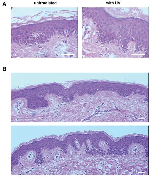
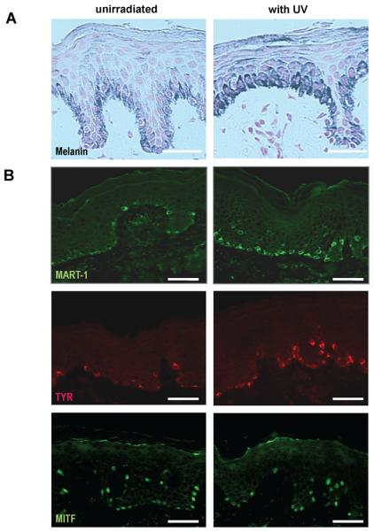
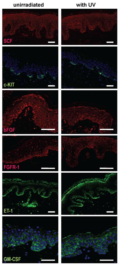
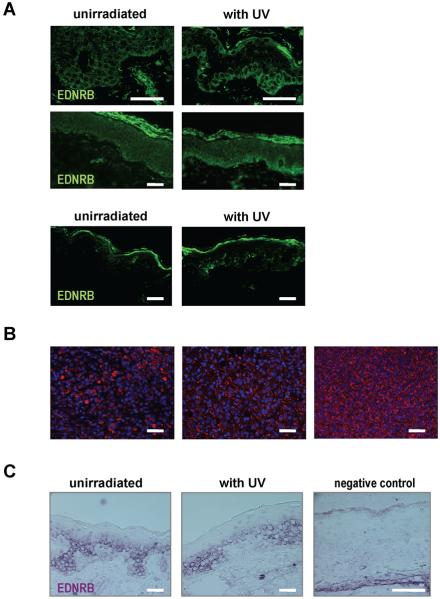
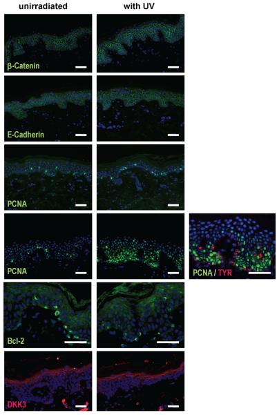
It is known that UV modulates the expression of paracrine factors that regulate melanocyte function in the skin. We investigated the consequences of repetitive UV exposure of human skin in biopsies of 10 subjects with phototypes 2-3.5 taken 1-4 years later. The expression of melanogenic factors (TYR, MART1, MITF), growth factors/receptors (SCF/KIT, bFGF/FGFR1, ET1/EDNRB, HGF, GM-CSF), adhesion molecules (beta-catenin, E-cadherin, N-cadherin), cell cycle proteins (PCNA, cyclins D1, E2) as well as Bcl-2, DKK1, and DKK3, were analyzed by immunohistochemistry. Most of those markers showed no detectable changes at > or = 1 year after the repetitive UV irradiation. Although increased expression of EDNRB protein was detected in 3 of 10 UV-irradiated subjects, there was no detectable change in the expression of ET1 protein or in EDNRB mRNA levels. In summary, only the expression of TYR, MART1, and/or EDNRB, and only in some subjects, was elevated at > or = 1 year after UV irradiation. Thus the long-term effects of repetitive UV irradiation on human skin did not lead to significant changes in skin morphology and there is considerable subject-to-subject variation in responses. The possibility that changes in the expression and function of EDNRB triggers downstream activation of abnormal melanocyte proliferation and differentiation deserves further investigation.
Figures

References
-
- Ahn GY, Butt KI, Jindo T, Yaguchi H, Tsuboi R, Ogawa H. The expression of endothelin-1 and its binding sites in mouse skin increased after ultraviolet B irradiation or local injection of tumor necrosis factor alpha. J Dermatol. 1998;25:78–84. - PubMed
-
- Aoki H, Moro O, Tagami H, Kishimoto J. Gene expression profiling analysis of solar lentigo in relation to immunohistochemical characteristics. Br J Dermatol. 2007;156:1214–1223. - PubMed
-
- Bancroft JD, Stevens A. Theory and Practice of Histological Techniques. Churchill Livingstone; New York: 1982.
-
- Berking C, Takemoto R, Satyamoorthy K, Shirakawa T, Eskandarpour M, Hansson J, VanBelle PA, Elder DE, Herlyn M. Induction of melanoma phenotypes in human skin by growth factors and ultraviolet B. Cancer Res. 2004;64:807–811. - PubMed
-
- Bittner M, Meltzer P, Chen Y, Jiang Y, Seftor E, Hendrix M, Radmacher M, Simon R, Yakhini Z, Ben-Dor A, Sampas N, Dougherty E, Wang E, Marincola F, Gooden C, Lueders J, Glatfelter A, Pollock P, Carpten J, Gillanders E, Leja D, Dietrich K, Beaudry C, Berens M, Alberts D, Sondak V. Molecular classification of cutaneous malignant melanoma by gene expression profiling. Nature. 2000;406:536–540. - PubMed
Publication types
MeSH terms
Substances
Grants and funding
LinkOut - more resources
Full Text Sources
Research Materials
Miscellaneous
