Comparative Study
. 2008;3(10):e3497.
doi: 10.1371/journal.pone.0003497. Epub 2008 Oct 23.

Adherent monomer-misfolded SOD1

Affiliations
Comparative Study

Adherent monomer-misfolded SOD1

Yasuhiro Watanabe et al. PLoS One. 2008.

Abstract

Background: Multiple cellular functions are compromised in amyotrophic lateral sclerosis (ALS). In familial ALS (FALS) with Cu/Zn superoxide dismutase (SOD1) mutations, the mechanisms by which the mutation in SOD1 leads to such a wide range of abnormalities remains elusive.

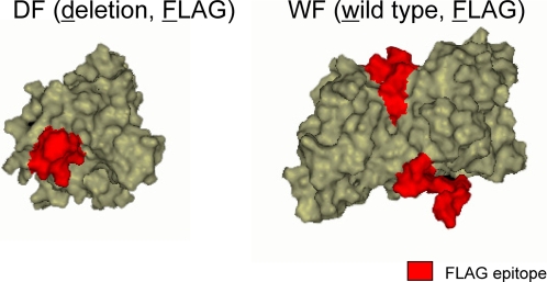
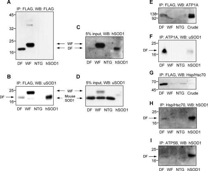
Methodology/principal findings: To investigate underlying cellular conditions caused by the SOD1 mutation, we explored mutant SOD1-interacting proteins in the spinal cord of symptomatic transgenic mice expressing a mutant SOD1, SOD1(Leu126delTT) with a FLAG sequence (DF mice). This gene product is structurally unable to form a functional homodimer. Tissues were obtained from both DF mice and disease-free mice expressing wild-type with FLAG SOD1 (WF mice). Both FLAG-tagged SOD1 and cross-linking proteins were enriched and subjected to a shotgun proteomic analysis. We identified 34 proteins (or protein subunits) in DF preparations, while in WF preparations, interactions were detected with only 4 proteins.

Conclusions/significance: These results indicate that disease-causing mutant SOD1 likely leads to inadequate protein-protein interactions. This could be an early and crucial process in the pathogenesis of FALS.


Conflict of interest statement

Competing Interests: The authors have declared that no competing interests exist.

Figures

Figure 1
Figure 1. Computer assisted simulation of the FLAG-tagged SOD1 proteins.
Simulation models of the FLAG-tagged SOD1s were constituted using Discovery Studio 1.7 software (Accelrys, USA) from Protein Data Bank (PDB) data; 1MFM for DF, 2V0A for WF as templates. The red parts indicate the FLAG epitopes. The DF mutant SOD1 possibly exposes a hydrophobic structure, which is normally buried by the dimer interface.
Figure 2
Figure 2. Immunoprecipitation and western blot analyses of mouse spinal cords.
The FLAG-tagged SOD1 and the ligand proteins were immunoprecipitated using the FLAG affinity resin and detected by the anti FLAG antibody (A) and anti-SOD1 polyclonal antibody (uSOD1) that recognizes both human and mouse SOD1 (B). The DF mutant SOD1 shows a distinct single band (A and B). In contrast, the WF shows high molecular weight bands (A) and a low molecular weight band which is possibly the mouse intrinsic SOD1 (B), indicating that the WF forms the heterodimer with the mouse SOD1. Crude spinal cord extracts (5% input) were analyzed using antibodies for human specific SOD1 (hSOD1) (C) and for uSOD1 (D). Compared to the WF, the amount of the DF SOD1 protein is considerably lower (C). The WF itself is lower still compared to the mouse intrinsic SOD1 (D). The immunoprecipitation products with the FLAG were analyzed by antibodies for ATP1A (E) and Hsp/Hsc70 (G). In this analysis, only the DF preparations show the presence of ATP1A (E). In the Hsp/Hsc70 analysis in (G), there are non-specific precipitations also in WF and NTG. In contrast, the immunoprecipitated samples for ATP1A (F), Hsp/Hsc70 (H) and ATP5B (I) were analyzed using antibodies for hSOD1 (F) or uSOD1 (H and I). Correspondingly, the human form of SOD1 is only seen to be co-immunoprecipitated in DF preparation (F, H and I). Lanes from the left are: DF, WF, NTG, and a purified human SOD1 (A, B, C, D, F, H, and I); and DF, WF, NTG, and a crude soluble fraction of NTG mice (E and G).
Figure 3
Figure 3. Immunohistochemistry.
Fixed-frozen sections of the spinal cord (10 µm thickness) derived from symptomatic DF mice at 145 days of life (A, C, E, and G) and age-matched NTG mice (B, D, F, and H) were stained with antibodies for ATP1A (A and B), ATP1B (C and D), ATP5A (E and F), ATP5B (G and H). The tissue was counterstained with hematoxylin. Bar: 200 µm.

References

    1. Cleveland DW, Rothstein JD. From Charcot to Lou Gehrig: deciphering selective motor neuron death in ALS. Nat Rev Neurosci. 2001;2:806–819. - PubMed
    1. Pasinelli P, Brown RH. Molecular biology of amyotrophic lateral sclerosis: insights from genetics. Nat Rev Neurosci. 2006;7:710–723. - PubMed
    1. Rosen DR, Siddique T, Patterson D, Figlewicz DA, Sapp P, et al. Mutations in Cu/Zn superoxide dismutase gene are associated with familial amyotrophic lateral sclerosis. Nature. 1993;362:59–62. - PubMed
    1. Boillee S, Yamanaka K, Lobsiger CS, Copeland NG, Jenkins NA, et al. Onset and progression in inherited ALS determined by motor neurons and microglia. Science. 2006;312:1389–1392. - PubMed
    1. Takahashi K, Nakamura H, Okada E. Hereditary amyotrophic lateral sclerosis. Histochemical and electron microscopic study of hyaline inclusions in motor neurons. Arch Neurol. 1972;27:292–299. - PubMed

Publication types

MeSH terms