. 2009 Apr;91(4):283-96.
doi: 10.1002/bip.21107.

Noncovalent probes for the investigation of structure and dynamics of protein-nucleic acid assemblies: the case of NC-mediated dimerization of genomic RNA in HIV-1

Affiliations

Noncovalent probes for the investigation of structure and dynamics of protein-nucleic acid assemblies: the case of NC-mediated dimerization of genomic RNA in HIV-1

Kevin B Turner et al. Biopolymers. 2009 Apr.

Abstract

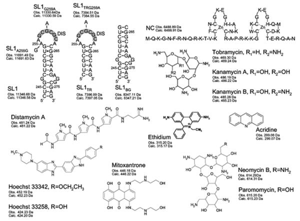
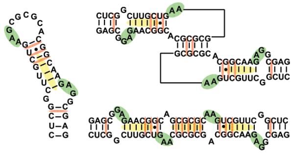
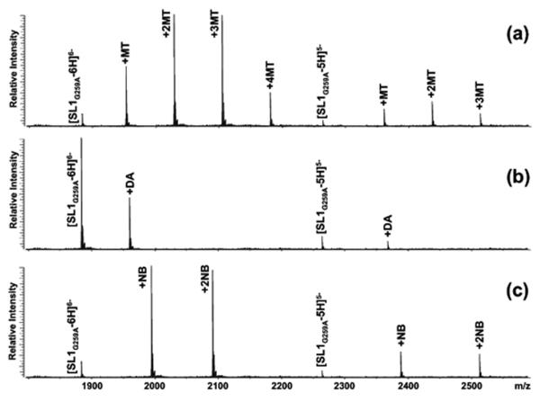
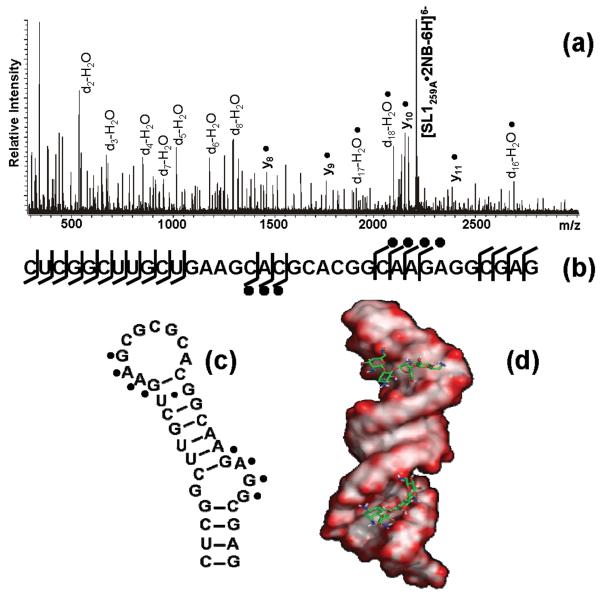
The nature of specific RNA-RNA and protein-RNA interactions involved in the process of genome dimerization and isomerization in HIV-1, which is mediated in vitro by stemloop 1 (SL1) of the packaging signal and by the nucleocapsid (NC) domain of the viral Gag polyprotein, was investigated by using archetypical nucleic acid ligands as noncovalent probes. Small-molecule ligands make contact with their target substrates through complex combinations of H-bonds, salt bridges, and hydrophobic interactions. Therefore, their binding patterns assessed by electrospray ionization mass spectrometry can provide valuable insights into the factors determining specific recognition between species involved in biopolymer assemblies. In the case of SL1, dimerization and isomerization create unique structural features capable of sustaining stable interactions with classic nucleic acid ligands. The binding modes exhibited by intercalators and minor groove binders were adversely affected by the significant distortion of the duplex formed by palindrome annealing in the kissing-loop (KL) dimer, whereas the modes observed for the corresponding extended duplex (ED) confirmed a more regular helical structure. Consistent with the ability to establish electrostatic interactions with highly negative pockets typical of helix anomalies, polycationic aminoglycosides bound to the stem-bulge motif conserved in all SL1 conformers, to the unpaired nucleotides located at the hinge between kissing hairpins in KL, and to the exposed bases flanking the palindrome duplex in ED. The patterns afforded by intercalators and minor groove binders did not display detectable variations when the corresponding NC-SL1 complexes were submitted to probing. In contrast, aminoglycosides displayed the ability to compete with the protein for overlapping sites, producing opposite effects on the isomerization process. Indeed, displacing NC from the stem-bulges of the KL dimer induced inhibition of stem melting and decreased the efficiency of isomerization. Competition for the hinge region, instead, eliminated the NC stabilization of a grip motif formed by nucleobases of opposite strands, thus facilitating the strand-exchange required for isomerization. These noncovalent probes provided further evidence that the structural context of the actual binding sites has significant influence on the chaperone activities of NC, which should be taken in account when developing potential drug candidates aimed at disrupting genome dimerization and isomerization in HIV-1.


Figures

Scheme 1
Scheme 1
Sequences and secondary structures of the ribonucleic acid constructs and nucleocapsid protein included in the study. Nucleotides are numbered according to the subtype B sequence (Lai variant) of HIV-1. The self-complementary sequences are highlighted in gray. For each species, the monoisotopic masses observed experimentally and calculated from sequence are included. Structures of nucleic acid-active agents incorporated in the study, calculated from monoisotopic masses and experimentally determined by FTICR.
Scheme 2
Scheme 2
Sequences and secondary structures of the monomer, kissing-loop, and extended duplex conformations of SL1. Shaded regions indicate proposed binding sites of the aminoglycosides (green), intercalators (red), and minor-groove binders (yellow).
Figure 1
Figure 1
Nanospray-FTICR mass spectra of 5 μM solutions of dimerization-deficient SL1G259A in 150mM ammonium acetate after a 5-fold addition of: (a) mitoxantrone; (b) distamycin A; (c) neomycin B (see Materials and Methods).
Figure 2
Figure 2
(a) Product ion spectrum and (b) fragmentation map of the SL1G259A•2NB assembly. The fragmentation pattern obtained by SORI-CID displayed protection of G254:G257 and G270:G272, which are located in double-stranded regions near the internal bulge and immediately below the apical loop, as indicated on the hairpin secondary structure (c). These results were corroborated by docking the ligand molecule onto the electrostatic surfaces of SL1 (d). Docking simulations were performed using Autodock 3.0 [67] (see Materials and Methods), using electrostatic surfaces calculated by Delphi [68] from the high-resolution coordinates of SL1 (PDB:2D1B) in a 150 mM salt environment. The results were visualized in Pymol [69], with red color marking highly electronegative regions.
Figure 3
Figure 3
Product ion spectra obtained by submitting to mild SORI-CID (a) the 2SL1•10MT complex folded via the KL-inducing protocol and (b) the 2SL1•13MT folded via the ED-inducing protocol. The former results in the dissociation of the dimer coupled with the loss of two mitoxantrone units, while the latter remains intact under the same energy regime. The precursor ion is marked by the formula image symbol.
Figure 4
Figure 4
Nanospray-FTICR mass spectra of a 5 μM solution of 2NC•SL1 G259A assembly in 150mM ammonium acetate after addition of 5-fold excess of (a) mitoxantrone, (b) distamycin A, and (c) neomycin B. The latter induced dissociation of the 2NC•SL1 G259A complex, while the two former classes of ligands create ternary complexes with 444.20 and 481.24 Da increments, respectively.
Figure 5
Figure 5
Nanospray-FTICR mass spectrum of a sample containing 30 μM NC (○) and 10 μM SL1A255G (◆) in 150 mM ammonium acetate (pH 7.0) after 3 hour incubation at 37°C in the (a) absence of ligand, (b) in the presence of 30 μM and (c) 50μM neomycin B (•). The percentages in parentheses indicate the proportions of KL/ED within each protein-RNA assembly obtained by tandem mass spectrometry under mild activation conditions (see Materials and Methods). From the resulting spectra, dimeric products were assigned to the ED conformer, whereas monomeric products were assigned to KL (as shown in Figure 3). The percentages were then summed to determine the partitioning between the two conformers within each precursor ion population, as reported in the box [38].

Similar articles

Cited by

References

    1. Peattie DA, Gilbert W. Chemical probes for higher-order structure in RNA. Proc. Natl. Acad. Sci. U.S.A. 1980;77(8):4679–82. - PMC - PubMed
    1. Walker TA, et al. Enzymatic and chemical structure mapping of mouse 28S ribosomal ribonucleic acid contacts in 5.8S ribosomal ribonucleic acid. Biochemistry. 1982;21:2320–29. - PubMed
    1. Ehresmann C, et al. Probing the structure of RNAs in solution. Nucleic Acids Res. 1987;12(22):9109–9128. - PMC - PubMed
    1. Krol A, Carbon P. A guide for probing native small nuclear RNA and ribonucleoprotein structures. Methods Enzymol. 1989;180:212–27. - PubMed
    1. Brunel C, Romby P. Probing RNA structure and RNA-ligand complexes with chemical probes. Methods Enzymol. 2000;318:3–21. - PubMed

Publication types

LinkOut - more resources