. 2008 Oct 23:5:48.
doi: 10.1186/1742-2094-5-48.

Neurological and behavioral abnormalities, ventricular dilatation, altered cellular functions, inflammation, and neuronal injury in brains of mice due to common, persistent, parasitic infection

Affiliations

Neurological and behavioral abnormalities, ventricular dilatation, altered cellular functions, inflammation, and neuronal injury in brains of mice due to common, persistent, parasitic infection

Gretchen Hermes et al. J Neuroinflammation. .

Abstract

Background: Worldwide, approximately two billion people are chronically infected with Toxoplasma gondii with largely unknown consequences.

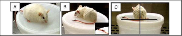
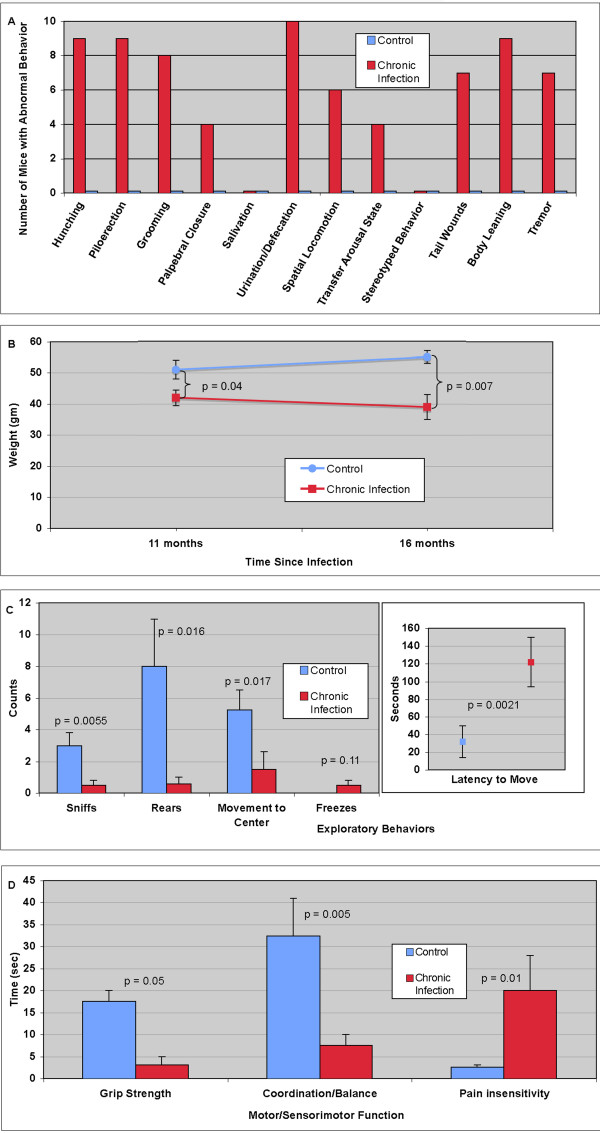
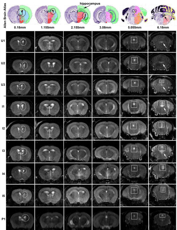
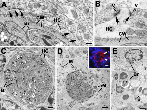
Methods: To better understand long-term effects and pathogenesis of this common, persistent brain infection, mice were infected at a time in human years equivalent to early to mid adulthood and studied 5-12 months later. Appearance, behavior, neurologic function and brain MRIs were studied. Additional analyses of pathogenesis included: correlation of brain weight and neurologic findings; histopathology focusing on brain regions; full genome microarrays; immunohistochemistry characterizing inflammatory cells; determination of presence of tachyzoites and bradyzoites; electron microscopy; and study of markers of inflammation in serum. Histopathology in genetically resistant mice and cytokine and NRAMP knockout mice, effects of inoculation of isolated parasites, and treatment with sulfadiazine or alphaPD1 ligand were studied.

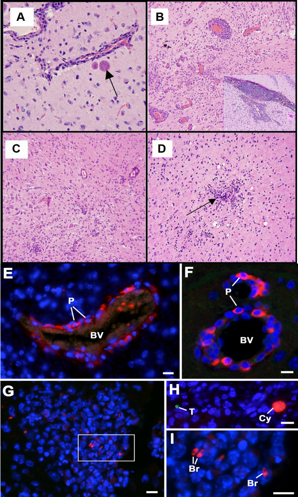
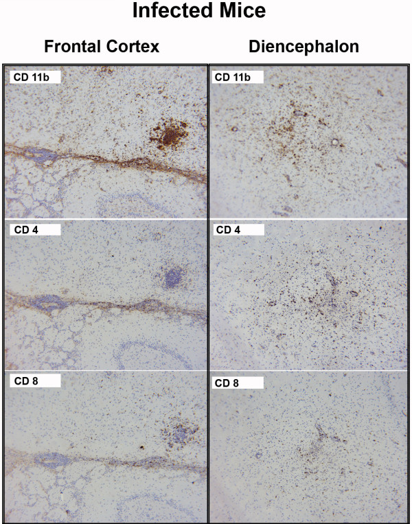
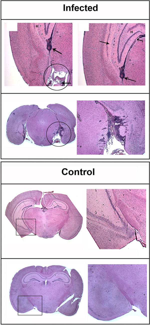
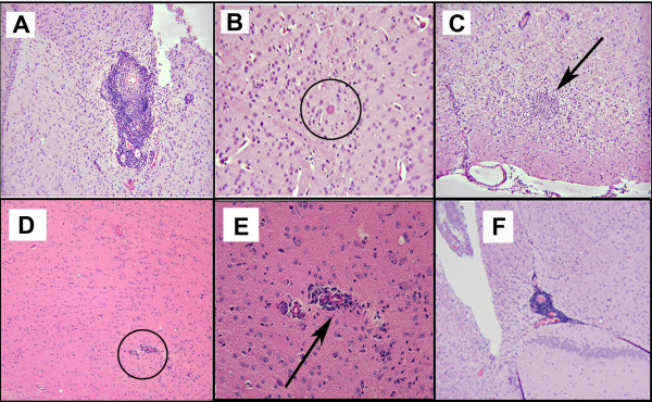
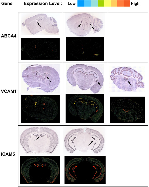
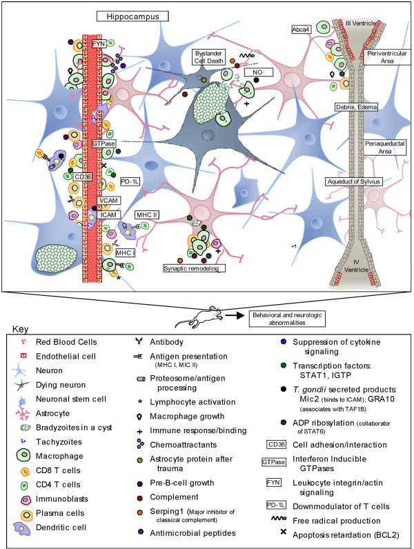
Results: Twelve months after infection, a time equivalent to middle to early elderly ages, mice had behavioral and neurological deficits, and brain MRIs showed mild to moderate ventricular dilatation. Lower brain weight correlated with greater magnitude of neurologic abnormalities and inflammation. Full genome microarrays of brains reflected inflammation causing neuronal damage (Gfap), effects on host cell protein processing (ubiquitin ligase), synapse remodeling (Complement 1q), and also increased expression of PD-1L (a ligand that allows persistent LCMV brain infection) and CD 36 (a fatty acid translocase and oxidized LDL receptor that mediates innate immune response to beta amyloid which is associated with pro-inflammation in Alzheimer's disease). Immunostaining detected no inflammation around intra-neuronal cysts, practically no free tachyzoites, and only rare bradyzoites. Nonetheless, there were perivascular, leptomeningeal inflammatory cells, particularly contiguous to the aqueduct of Sylvius and hippocampus, CD4+ and CD8+ T cells, and activated microglia in perivascular areas and brain parenchyma. Genetically resistant, chronically infected mice had substantially less inflammation.

Conclusion: In outbred mice, chronic, adult acquired T. gondii infection causes neurologic and behavioral abnormalities secondary to inflammation and loss of brain parenchyma. Perivascular inflammation is prominent particularly contiguous to the aqueduct of Sylvius and hippocampus. Even resistant mice have perivascular inflammation. This mouse model of chronic T. gondii infection raises questions of whether persistence of this parasite in brain can cause inflammation or neurodegeneration in genetically susceptible hosts.


Figures

Figure 1
Figure 1
Appearance of eleven-month-old Specific Pathogen Free (SPF) mice that are uninfected and chronically infected with T. gondii. (A) Eleven month old uninfected Swiss Webster mouse. (B) Eleven month old infected female mouse from the same SPF colony ten months after acquisition of T. gondii infection. Note hunched body position, poor grooming, piloerection, reduced body mass, and tail wounding. (C) Chronically infected SPF mouse with abnormal body position.
Figure 2
Figure 2
Behavioral and Neurologic Data of eleven-month-old Specific Pathogen Free (SPF) mice that are uninfected and chronically infected with T. gondii. (A) Number of mice with abnormal behavior or neurologic findings. (B) Weight of uninfected and chronically infected mice at eleven (p = 0.04) and sixteen months (p = 0.007) after infection. Error bars indicate one standard error. (C) Exploratory behaviors in mice twelve months after infection. Error bars indicate one standard error. (D) Motor and sensorimotor function in uninfected and infected mice eleven to sixteen months after infection. Error bars indicate one standard error. n = 6 control; n = 10 infected in all cases.
Figure 3
Figure 3
MRI findings in chronically infected mice and in uninfected mice. T2 weighted MRIs from uninfected mice (U1-3), chronically infected mice (I1-5), and one infected mouse studied in a pilot experiment at a different time (P1). MRIs were obtained when mice were 1 year old and infected mice had been infected for 8 months. In infected mice there was minimal to moderate ventricular dilation. Images from the Allen Brain Atlas are provided in the top panel for reference to the brain region imaged. Lateral ventricles are marked with a circle and the 3rd ventricle/aqueduct of Sylvius is marked with a square. Unless otherwise noted, the 4th ventricle is marked with an arrow. T2 weighted MRI of the brain in infected and age-matched control mice show an increase of size in the lateral ventricle of infected mice (p = 0.034 approximately 1.155 mm caudal to bregma) and an increase in 3rd ventricle size (marked by a square) of infected mice (far right panel, p = 0.002). In 4 of 6 infected mice there is right-left brain asymmetry (I1, I3, I4, I5). The major difference between the infected and the uninfected MRIs was ventricular dilatation and some asymmetry present in brain parenchyma of infected mice.
Figure 4
Figure 4
Inflammation in the brain during chronic Toxoplasma infection with only a few bradyzoites and cysts. (A) Occasional cyst with bradyzoites a short distance from a vessel (arrow), × 250. (B) Medium power view showing perivascular cuffing, × 100. (C) High power view of brain. Note perivascular cuffing and microglial infiltrates. Meningeal infiltrates also occurred (not shown), × 100. (D) Small numbers of microglial nodules (arrow) with diffuse inflammatory infiltrates throughout brain parenchyma, × 100. (E-I) Sections through the brains of chronically infected mice from the behavioral and neurologic studies, double labeled with rabbit anti-SAG1 visualised with anti-rabbit Ig conjugated to fluorescein isothiocyanate (green) and mouse anti-BAG1 visualised with anti-mouse Ig conjugated to Texas red (red). Bars represent 10 μm. (E, F) Examples of blood vessels (BV) cuffed with numerous inflammatory cells in which the plasma cells (P) can be identified by the cross reaction of the anti-mouse Ig with the immunoglobulins within the plasma cell cytoplasm (red), × 400. (G) Low power of a nodule of inflammatory cells in which a few bradyzoites (red) but no tachyzoites (green) could be identified, × 100. (H) A section of brain showing a single tachyzoite (green) and tissue cyst (red) in an area with no inflammatory cells, × 100. (I) Detail from the enclosed area in G showing the cytoplasmic staining of the bradyzoites (Br) with anti-BAG1, × 1000.
Figure 5
Figure 5
T cells and microglia in brains of chronically infected mice. Representative images of one of five infected mice. T lymphocytes were present in nodules and in the perivascular spaces in frontal cortex and diencephalons. CD4 T lymphocytes and microglia and CD8 T lymphocytes ×40. Immunohistochemical stains for CD11 indicate activated microglia. × 100. H & E stains also were performed. In contrast, preparations of brains from 5 uninfected mice revealed no inflammatory cells other than occasional microglial cells (not shown). Secondary antibody control stained tissues had no background staining (not shown).
Figure 6
Figure 6
Perivascular inflammatory infiltrates in vessels that supply the hippocampus (circle) adjacent to the hippocampus (labeled H) and in vessels contiguous to and in the hippocampus (arrows) and at the base of the brain. No such inflammatory cells are seen in uninfected control mice.
Figure 7
Figure 7
(A-C) Similar histopathology with perivascular inflammation, isolated cyst ×40, and cluster of microglia in a mouse that is chronically infected, initially infected with parasites without accompanying brain. A ×40; B ×250; C ×20. (D) Less prominent perivascular cuffing (circle) and small collections of mononuclear cells in Balb/C mouse that is genetically more resistant. E. Increased magnification of area of perivascular inflammation. × 100.(F) Perivascular inflammation × 40 for mouse that had mild lateral ventricular dilatation in MRI.
Figure 8
Figure 8
(A-B) Details of the periphery of tissue cysts from chronically infected mice identifying the host cells (HC) as neurons due to the formation of synapses (arrows). Based on the structure of the synapse the cysts appear to be within a neuronal dendrite. V – neurosecretory vescicles; CW – cyst wall; Br – bradyzoite. Bars represent 100 nm. (C) Low power of a cyst containing numerous bradyzoites (Br) within a host cell (HC). Note the absence of inflammatory cells. Bar is 1 μm. (D) Low power of a ruptured tissue cyst showing numerous inflammatory cells (M) around and invading through breaks in the cyst wall into the tissue cyst. Bar is 10 μm. Inset: Similar area to that in D immunostained with an antibody to the cyst wall (CC2) showing the disruption to the tissue cyst wall (arrows) and the surrounding inflammatory cells. Bar is 10 μm. (E) Detail from D showing bradyzoites (Br) that had been phagocytised by inflammatory cells. Bar is 1 μm.
Figure 9
Figure 9
ABCA4, VCAM and ICAM are expressed in the perihippocampal, hippocampal and periventricular areas. Images obtained from the Allen Brain Atlas.
Figure 10
Figure 10
Schematic summary of cells, molecules, and processes affected by chronic T. gondii infection resulting in neuronal cell loss, inflammation, and behavioral and other neurologic abnormalities in mice. Parasites initially arrive at blood brain barrier in dendritic cells, travel into brain tissue and, within neurons, form bradyzoites in cysts. When the neuronal membrane is no longer intact, bradyzoites elicit an immune response as shown, which results in destruction of their growth and bystander cell death as shown.

References

    1. Boyer K, Marcinak J, McLeod R. Toxoplasma gondii (Toxoplasmosis) In: Long S, Pickering LK, Prober CG, editor. Principles and Practice of Pediatric Infectious Diseases. 3. New York: Churchill Livingstone; 2007.
    1. Mortensen PB, Norgaard-Pedersen B, Waltoft BL, Sorensen TL, Hougaard D, Yolken RH. Early infections of Toxoplasma gondii and the later development of schizophrenia. Schizophr Bull. 2007;33:741–744. - PMC - PubMed
    1. Flegr J, Preiss M, Klose J, Havlicek J, Vitakova M, Kodym P. Decreased level of psychobiological factor novelty seeking and lower intelligence in men latently infected with the protozoan parasite Toxoplasma gondii Dopamine, a missing link between schizophrenia and toxoplasmosis? Biol Psychol. 2003;63:253–268. - PubMed
    1. Palmer BS. Meta-analysis of three case controlled studies and an ecological study into the link between cryptogenic epilepsy and chronic toxoplasmosis infection. Seizure. 2007;16:657–663. - PubMed
    1. Torrey EF, Bartko JJ, Lun ZR, Yolken RH. Antibodies to Toxoplasma gondii in patients with schizophrenia: a meta-analysis. Schizophr Bull. 2007;33:729–736. - PMC - PubMed

Publication types

MeSH terms