. 2008 Oct 28;18(20):1576-80.
doi: 10.1016/j.cub.2008.08.068.

fMRI adaptation reveals mirror neurons in human inferior parietal cortex

Affiliations

fMRI adaptation reveals mirror neurons in human inferior parietal cortex

Trevor T-J Chong et al. Curr Biol. .

Abstract

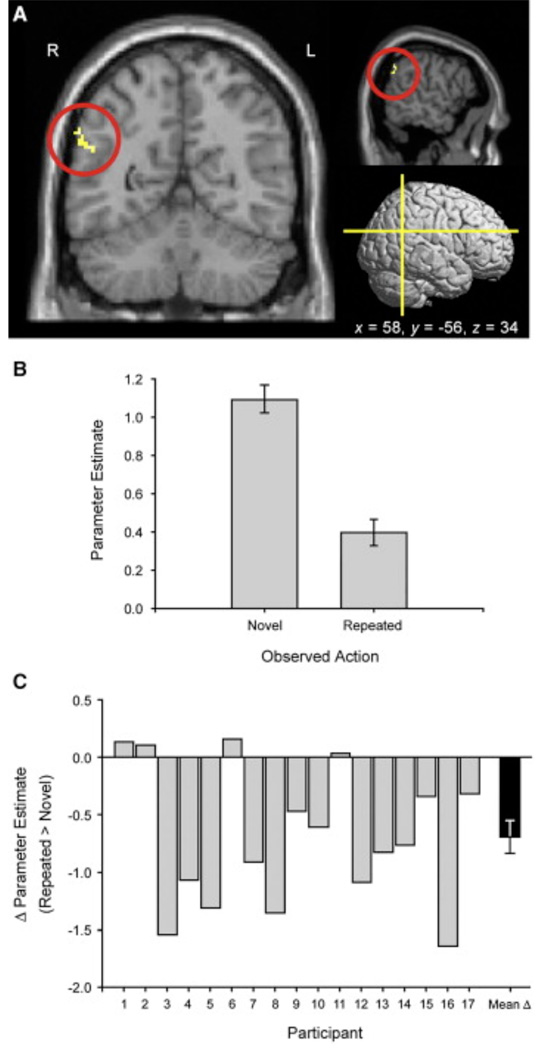
Mirror neurons, as originally described in the macaque, have two defining properties [1, 2]: They respond specifically to a particular action (e.g., bringing an object to the mouth), and they produce their action-specific responses independent of whether the monkey executes the action or passively observes a conspecific performing the same action. In humans, action observation and action execution engage a network of frontal, parietal, and temporal areas. However, it is unclear whether these responses reflect the activity of a single population that represents both observed and executed actions in a common neural code or the activity of distinct but overlapping populations of exclusively perceptual and motor neurons [3]. Here, we used fMRI adaptation to show that the right inferior parietal lobe (IPL) responds independently to specific actions regardless of whether they are observed or executed. Specifically, responses in the right IPL were attenuated when participants observed a recently executed action relative to one that had not previously been performed. This adaptation across action and perception demonstrates that the right IPL responds selectively to the motoric and perceptual representations of actions and is the first evidence for a neural response in humans that shows both defining properties of mirror neurons.


Figures

Figure 1
Figure 1. Experimental Design of the fMRI Adaptation Paradigm in Experiment 1
(A) In the “Execute-Observe” runs, blocks comprised a series of “Execute” trials followed by an equivalent number of “Observe” trials. “Execute” trials required participants to commence a cued action at the onset of a central fixation dot. “Observe” trials required participants to passively observe a presented action. The observed actions were either the same as those previously executed (as illustrated), or different from them. (B) The “Observe-Execute” runs reversed the order of “Execute” and “Observe” trials.
Figure 2
Figure 2. Locus of fMRI Adaptation within the Right IPL during the “Execute-Observe” Runs of Experiment 1
(A) Coronal, sagittal, and rendered lateral views of the right IPL cluster, with its peak voxel at MNI 58, −56, 34, p < 0.001. (B) Parameter estimates for the Observe Novel and Observe Repeated trials across the right IPL cluster. (C) Magnitude of repetition suppression for the IPL cluster in single participants (gray bars) and across the group (black bar), expressed as the difference in parameter estimates between the observation of a Novel action and that of a Repeated action. Negative differences indicate significant repetition suppression. Error bars indicate ± 1 SEM. The data in (B) and (C) are derived from the statistical test (shown in [A]) by which the right IPL cluster was selected.
Figure 3
Figure 3. Experimental Design of the fMRI Adaptation Paradigm and Results for Experiment 2
(A) The event-related design consisted of sparsely presented pairs of “Execute” and “Observe” trials. The actions presented in the “Observe” trials were either identical to (as illustrated) or different from the previously executed action. (B) Magnitude of fMRI adaptation across the right IPL cluster defined in experiment 1, in single participants (gray bars) and across the group (black bar), expressed as the difference in parameter estimates between the observation of a Novel action and that of a Repeated action. Negative differences indicate significant adaptation. (C) Time course of the adaptation effect, shown as a plot of percent signal change against time. Error bars indicate ± 1 SEM. The data in (B) and (C) are independent of the data used to select these voxels and hence provide independent confirmation of the effect shown in Figure 2A.

Similar articles

Cited by

References

    1. Gallese V, Fadiga L, Fogassi L, Rizzolatti G. Action recognition in the premotor cortex. Brain. 1996;119:593–609. - PubMed
    1. Di Pellegrino G, Fadiga L, Fogassi L, Gallese V, Rizzolatti G. Understanding motor events: A neurophysiological study. Exp. Brain Res. 1992;91:176–180. - PubMed
    1. Dinstein I, Thomas C, Behrmann M, Heeger DJ. A mirror up to nature. Curr. Biol. 2008;18:R13–R18. - PMC - PubMed
    1. Rizzolatti G, Craighero L. The mirror-neuron system. Annu. Rev. Neurosci. 2004;27:169–192. - PubMed
    1. Fogassi L, Ferrari P, Gesierich B, Rozzi S, Chersi F, Rizzolatti G. Parietal lobe: From action organization to intention understanding. Science. 2005;308:662–667. - PubMed

Publication types

MeSH terms