. 2008 Dec 12;283(50):35129-39.
doi: 10.1074/jbc.M806008200. Epub 2008 Oct 23.

A novel protein domain induces high affinity selenocysteine insertion sequence binding and elongation factor recruitment

Affiliations

A novel protein domain induces high affinity selenocysteine insertion sequence binding and elongation factor recruitment

Jesse Donovan et al. J Biol Chem. .

Abstract

Selenocysteine (Sec) is incorporated at UGA codons in mRNAs possessing a Sec insertion sequence (SECIS) element in their 3'-untranslated region. At least three additional factors are necessary for Sec incorporation: SECIS-binding protein 2 (SBP2), Sec-tRNA(Sec), and a Sec-specific translation elongation factor (eEFSec). The C-terminal half of SBP2 is sufficient to promote Sec incorporation in vitro, which is carried out by the concerted action of a novel Sec incorporation domain and an L7Ae RNA-binding domain. Using alanine scanning mutagenesis, we show that two distinct regions of the Sec incorporation domain are required for Sec incorporation. Physical separation of the Sec incorporation and RNA-binding domains revealed that they are able to function in trans and established a novel role of the Sec incorporation domain in promoting SECIS and eEFSec binding to the SBP2 RNA-binding domain. We propose a model in which SECIS binding induces a conformational change in SBP2 that recruits eEFSec, which in concert with the Sec incorporation domain gains access to the ribosomal A site.


Figures

FIGURE 1.
FIGURE 1.
Alignment of the CT-SBP2 sequences. Alignment of human (Hs), rat (Rn), and chicken (Gg) SBP2 and the human SBP2 paralogue, SBP2-like protein (Hs SLP) is shown. Residues mutated to alanine in groups of five (boxed) are indicated above the alignment. The SBP2 arrow also indicates the stop and start positions of the recombinant SID and RBD, respectively.
FIGURE 2.
FIGURE 2.
A, [35S]Met-labeled in vitro translated wild-type and mutant CT-SBP2 proteins were added to the in vitro translation of a Sec incorporation luciferase reporter containing an in-frame UGA codon and a GPX4 SECIS element in the 3′-untranslated region. B, graphical representation of the Sec incorporation activity expressed as the percentage of luciferase activity observed relative to wild-type CT-SBP2 (means ± S.E.).
FIGURE 3.
FIGURE 3.
Analysis of SECIS and ribosome binding for penta-alanine mutants. A, 16 fmol of [35S]Met labeled in vitro translated wild-type or mutant CT-SBP2, as indicated, was incubated with 32P-labeled wild-type GPX4 SECIS elements and resolved on 4% nondenaturing polyacrylamide gels (the last four mutants were evaluated in a separate experiment). B, [35S]Met-labeled in vitro translated CT-SBP2 was assayed for ribosome binding by pelleting complexes through a 20% sucrose cushion. Five percent of supernatants (S) and pellets (P) were resolved by SDS-PAGE (top panel) and visualized by PhosphorImaging. To control for nonspecific binding, the amount of pelleting observed for hnRNP F was subtracted and data were normalized to CT-SBP2. C, summary of Sec incorporation (black bars), ribosome binding (light gray bars), and SECIS binding (dark gray bars) data for the penta-alanine mutants expressed as the amount of each activity relative to that obtained with wild-type CT-SBP2 (means ± S.E., n = 3).
FIGURE 4.
FIGURE 4.
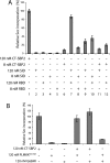
The domains of CT-SBP2 function in trans. A, the Sec incorporation luciferase reporter was translated in RRL in the presence of the indicated recombinant proteins. Luciferase activity is expressed as percentages of that obtained with wild-type CT-SBP2 (means ± S.E., n = 3). B, the same luciferase reporter was translated in RRL with the indicated recombinant proteins. The 504PLMKK508 penta-alanine mutant and G669R are in a CT-SBP2 background.
FIGURE 5.
FIGURE 5.
The SECIS element promotes formation of a stable SID-RBD complex. Wild-type or penta-alanine mutant recombinant XH-SID (SID) was incubated with equimolar amounts of FLAG-RBD and 32P-trace-labeled wild-type (wt) or mutant (mt) SECIS elements. The proteins in the supernatant (top panel) and pellet (middle panel) fractions were resolved by SDS-PAGE and detected by Western blot using an anti-SBP2 antibody. Fifty percent of the supernatants from SECIS containing reactions were removed, and RNA was extracted, precipitated, and resolved on a 6% denaturing gel (bottom panel). Lanes 4 and 5 of the bottom panel show the starting RNA.
FIGURE 6.
FIGURE 6.
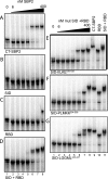
The SID enhances SECIS affinity. A-D, 2-fold serial dilutions of the recombinant proteins (6-400 nm) were incubated with 32P-labeled wild-type GPX4 SECIS elements, and complexes were resolved on 4% nondenaturing polyacrylamide gels and visualized by PhosphorImaging. E-G, same as D but with the SID mutant proteins indicated, and lanes 9-11 contain 400 nm each CT-SPB2, RBD, and SID + RBD, respectively.
FIGURE 7.
FIGURE 7.
The SID interacts transiently with the ribosome. A, [35S]Met-labeled in vitro translated proteins as indicated were assayed for ribosome binding as described for Fig. 3. The asterisk denotes a band that likely corresponds to the product of internal translation initiation at Met 629. B, [35S]Met-labeled in vitro translated proteins as indicated were cross-linked to the ribosome in 0.25% formaldehyde and spun through a 20% sucrose cushion containing 2% SDS. Amounts equal to 5% of supernatants and 20% of pellets were resolved by SDS-PAGE, visualized by PhosphorImaging, and quantitated using ImageQuant software as described for ribosome binding assays in Figs. 2 and 5.
FIGURE 8.
FIGURE 8.
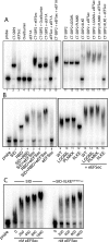
eEFSec forms a stable complex with SBP2. A, 32P-labeled wild-type GPX4 SECIS elements were incubated with 8 pmol of each of wild-type or mutant XH-CT-SBP2, FLAG-eEFSec, ovalbumin, or yeast eEF1A as indicated. B, same as in A except using 8 pmol each of XH-RBD, wild-type, or mutant XH-SID and FLAG-eEFSec as indicated. C, same as in A and B except using 8 pmol of wild-type or mutant XH-SID in the presence of increasing amounts of FLAG-eEFSec as indicated. In each case the lane labeled “probe” contains the wild-type 32P-labeled GPX4 SECIS element alone.
FIGURE 9.
FIGURE 9.
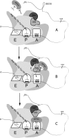
A model for eukaryotic Sec incorporation. An SBP2-bound ribosome encounters a UGA codon. At this step, prior to SECIS binding, eEFSec is blocked from the A site (A). A low affinity SECIS-RBD interaction induces a conformational change in the SID that generates a stable high affinity SECIS-RBD interaction in addition to SID-RBD interactions (not shown). The stable SID-RBD-SECIS complex is competent to recruit eEFSec to the ribosome (B). Once eEFSec has been recruited, we propose that a conformational change in the SID induces a conformation change in the ribosome that allows the eEFSec ternary complex to be fully accommodated in the ribosomal A site (B and C).

References

    1. Caban, K., and Copeland, P. R. (2006) Cell Mol. Life Sci. 63, 73-81 - PMC - PubMed
    1. Tujebajeva, R. M., Copeland, P. R., Xu, X. M., Carlson, B. A., Harney, J. W., Driscoll, D. M., Hatfield, D. L., and Berry, M. J. (2000) EMBO Rep. 1, 158-163 - PMC - PubMed
    1. Small-Howard, A., Morozova, N., Stoytcheva, Z., Forry, E. P., Mansell, J. B., Harney, J. W., Carlson, B. A., Xu, X. M., Hatfield, D. L., and Berry, M. J. (2006) Mol. Cell. Biol. 26, 2337-2346 - PMC - PubMed
    1. Zavacki, A. M., Mansell, J. B., Chung, M., Klimovitsky, B., Harney, J. W., and Berry, M. J. (2003) Mol Cell 11, 773-781 - PubMed
    1. Mehta, A., Rebsch, C. M., Kinzy, S. A., Fletcher, J. E., and Copeland, P. R. (2004) J. Biol. Chem. 279, 37852-37859 - PMC - PubMed

Publication types

LinkOut - more resources