. 2008 Dec;135(23):3959-68.
doi: 10.1242/dev.025304. Epub 2008 Oct 23.

The Mn1 transcription factor acts upstream of Tbx22 and preferentially regulates posterior palate growth in mice

Affiliations

The Mn1 transcription factor acts upstream of Tbx22 and preferentially regulates posterior palate growth in mice

Wenjin Liu et al. Development. 2008 Dec.

Abstract

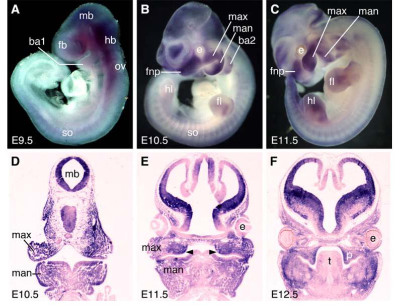
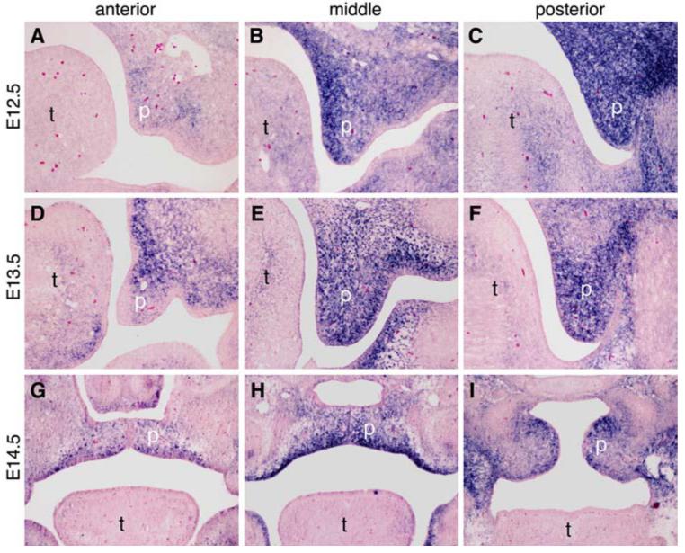
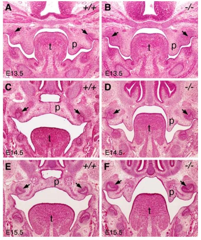
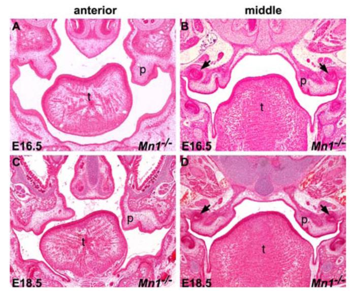
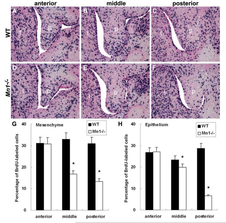
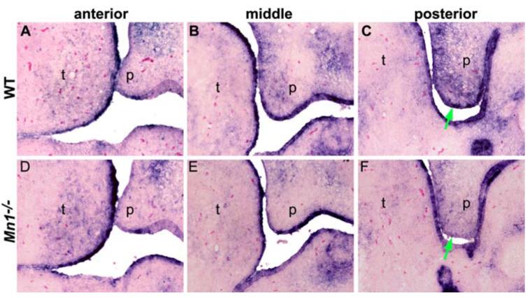
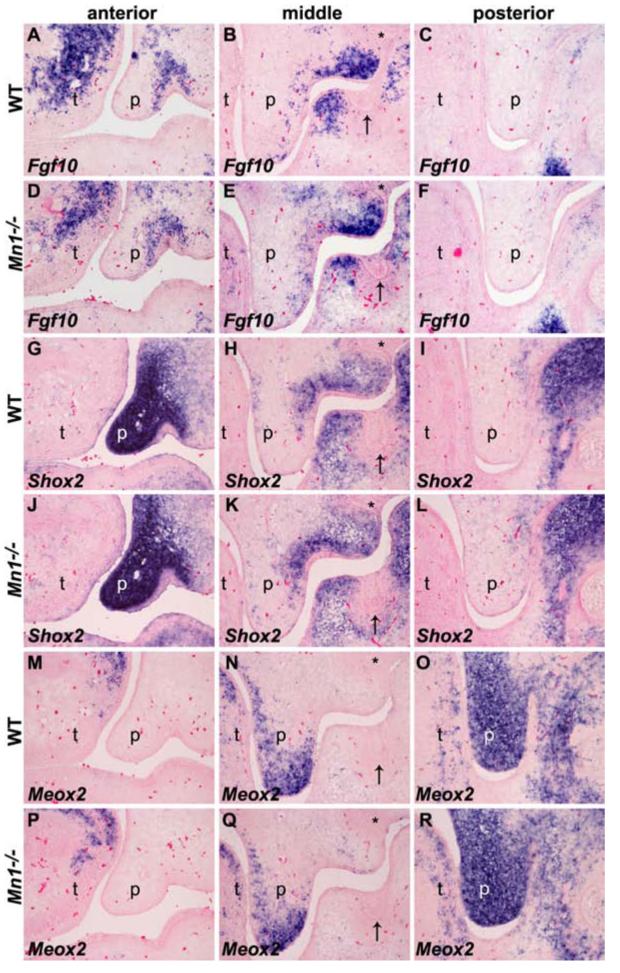
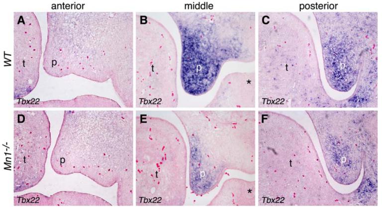
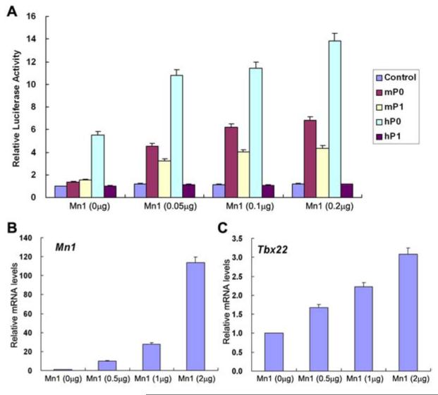
The mammalian secondary palate exhibits morphological, pathological and molecular heterogeneity along the anteroposterior axis. Although the cell proliferation rates are similar in the anterior and posterior regions during palatal outgrowth, previous studies have identified several signaling pathways and transcription factors that specifically regulate the growth of the anterior palate. By contrast, no factor has been shown to preferentially regulate posterior palatal growth. Here, we show that mice lacking the transcription factor Mn1 have defects in posterior but not anterior palatal growth. We show that Mn1 mRNA exhibits differential expression along the anteroposterior axis of the developing secondary palate, with preferential expression in the middle and posterior regions during palatal outgrowth. Extensive analyses of palatal gene expression in wild-type and Mn1(-/-) mutant mice identified Tbx22, the mouse homolog of the human X-linked cleft palate gene, as a putative downstream target of Mn1 transcriptional activation. Tbx22 exhibits a similar pattern of expression with that of Mn1 along the anteroposterior axis of the developing palatal shelves and its expression is specifically downregulated in Mn1(-/-) mutants. Moreover, we show that Mn1 activated reporter gene expression driven by either the human or mouse Tbx22 gene promoters in co-transfected NIH3T3 cells. Overexpression of Mn1 in NIH3T3 cells also increased endogenous Tbx22 mRNA expression in a dose-dependent manner. These data indicate that Mn1 and Tbx22 function in a novel molecular pathway regulating mammalian palate development.


Figures

Fig. 1
Fig. 1
Expression pattern of Mn1 mRNA in developing mouse embryos. mRNA signals were detected by whole mount in situ hybridization (A-C) or section in situ hybridization (D-F). (A) Mn1 mRNA expression was first detected in the developing brain tissues and in the craniofacial mesenchyme at E9.5. (B-E) Strong Mn1 mRNA expression was observed in the developing brain, frontonasal processes, maxillary processes, mandibular processes, the second branchial arch, the developing somites and limb buds at E10.5 (B, D) and E11.5 (C, E). (F) Section in situ hybridization showing strong Mn1 mRNA expression in the developing palatal mesenchyme and in the preossification mesenchymal cells in the mandible. ba1, first branchial arch; ba2, second branchial arch; e, eye; fb, forebrain; fl, forelimb; fnp, frontonasal process; hb, hindbrain; hl, hindlimb; man, mandibular process; max, maxillary process; mb, midbrain; ov, otic vesicle; p, palatal shelf; so, somite; t, tongue.
Fig. 2
Fig. 2
The expression pattern of Mn1 mRNA along the anterior-posterior axis of the developing palate. Middle palate corresponds to the palatal region flanked by the maxillary first molar tooth germs. (A-C) At E12.5, Mn1 mRNA expression in the developing secondary palate exhibits an anterior-posterior gradient, with low levels in the anterior palate (A), moderate levels in the middle palate (B), and highest levels in the posterior palate (C). (D-F) At E13.5, Mn1 mRNA expression is still high in the middle (E) and posterior (F) regions and much weaker in the anterior region (D) of the developing palate. (G-I) At E14.5, the bilateral palatal shelves have elevated to the horizontal position above the tongue and have initiated contact and fusion in the anterior (G) and middle (H) regions. Mn1 mRNA expression remains strong in the middle and posterior palatal regions and relatively weak in the anterior palate. p, palatal shelf; t, tongue.
Fig. 3
Fig. 3
Mn1-/- mutant mice exhibit palatal retardation and failure of palatal shelf elevation. All panels shown are from middle palate regions. (A, B) At E13.5, wild-type (A) and Mn1-/- homozygous mutant (B) embryos exhibited similar palatal shelf size and shape. (C, D) At E14.5, the wild-type (C) palatal shelves had elevated to the horizontal position above the tongue and initiated fusion by forming the midline epithelial seam, while the Mn1-/- homozygous mutant (D) palatal shelves were still vertically oriented. (E, F) At E15.5, the wild-type (E) palatal shelves had completed fusion, but the Mn1-/- homozygous mutant (F) palatal shelves remained vertically oriented. Arrows point to the first molar tooth germs. p, palatal shelf; t, tongue.
Fig. 4
Fig. 4
Histological analyses of palate development in Mn1-/- mutants at late stages. (A, B) At E16.5, the palatal shelves in Mn1-/- mutant embryos were still vertically oriented. In addition, the palatal shelves appeared to be significantly diminished in size in the middle to posterior regions (B). (C, D) At E18.5, whereas the palatal shelves in the anterior region in the Mn1-/- mutant mice were partially elevated (C), the middle and posterior regions of the palatal shelves were further retarded and retracted into the maxillary processes (D). Arrows in B and D point to the upper first molar tooth germs. p, palatal shelf; t, tongue.
Fig. 5
Fig. 5
Analyses of cell proliferation during palate development in wild-type and Mn1-/- mutant embryos. In comparison with the wild-type littermates (A-C), the number of BrdU-labeled cells in the developing palatal shelves was greatly reduced in the middle (E) and posterior (F) palatal shelves, but not in the anterior (D) palatal mesenchyme in the Mn1-/- mutant embryos by E13.5. p, palatal shelf; t, tongue. (G, H) Comparison of the percentage of BrdU-labeled cells in the palatal mesenchyme (G) and epithelium (H), respectively, in the anterior, middle, and posterior palatal regions in E13.5 wild-type (WT) and Mn1-/- mutant embryos. Error bars represent standard deviation and asterisk denotes a significant difference (P<0.01) between the wild-type and Mn1-/- mutant samples.
Fig. 6
Fig. 6
Analyses of cell proliferation and cell apoptosis in wild-type (WT) and Mn1-/- mutant embryos at E15.5. (A-C) Cell proliferation in palatal mesenchyme detected using immunohistochemical staining of the Ki67 protein. In comparison with wild-type embryos (A), cell proliferation in the middle palatal region in Mn1-/- mutant embryos (B) was greatly reduced. Error bars in (C) represent standard deviation and asterisk denotes a significant difference (P<0.01) between the wild-type and mutant samples. (D-E) TUNEL assays mid-palatal sections of E15.5 palatal shelves of wild-type (D) and Mn1-/- mutant (E) embryos showed increased cell apoptosis in the palatal mesenchyme in the mutant. Small arrows point to highly TUNEL-positive palatal mesenchymal cells and arrowheads point to highly TUNEL-positive palatal epithelial cells.
Fig. 7
Fig. 7
Expression of CyclinD2 was down-regulated in Mn1-/- mutant palatal shelves at E13.5. (A,D) In the anterior region of the developing palate, expression of CyclinD2 was similarly weak in wild-type (A) and Mn1-/- mutant (D) embryos. (B,E) In the middle palate, CyclinD2 is highly expressed in both the epithelium and mesenchyme in the wild-type embryo (B) but its expression is much reduced in the Mn1-/- palatal mesenchyme (E). (C) In the posterior palate region, strong CyclinD2 expression was detected in both the epithelium and mesenchyme in the wild-type palate. (F) In comparison to the wild-type littermate, CyclinD2 expression is much reduced in both the palatal epithelium and mesenchyme in the posterior palatal region in the Mn1-/- mutant embryo. Green arrows in C and F point to the medial edge epithelium of the palatal shelves.
Fig. 8
Fig. 8
The anterior-posterior pattern of the developing palatal shelves is not disrupted in the Mn1-/- mutant embryos. (A-F) Expression of Fgf10 along the anterior-posterior axis of the developing palatal shelves in wild-type (A-C) and Mn1-/- mutant (D-F) embryos at E13.5. Fgf10 showed similar restricted expression pattern in the anterior and middle palatal regions in both wild-type and Mn1-/- mutant embryos. (G-L) Expression of Shox2 along the anterior-posterior axis of the developing palatal shelves in wild-type (G-I) and Mn1-/- mutant (J-L) embryos at E13.5. Shox2 mRNA is strongly expressed in the anterior and absent in the posterior palatal regions in wild-type embryos (G-I). In the middle palate, Shox2 mRNA expression is restricted in the proximolateral region (H). The expression pattern and levels of Shox2 mRNA are not altered in Mn1-/- mutant (J-L) embryos. (M-O) Meox2 mRNA expression is restricted to the middle and posterior regions and absent from the anterior region of the developing palate in wild-type embryos at E13.5. (P-R) The levels and anterior-posterior pattern of Meox2 mRNA expression pattern in E13.5 Mn1-/- mutant embryo are comparable to that of the wild-type littermate (M-O). p, palatal shelf; t, tongue. Arrows point to the mandibular first molar tooth germs and asterisks mark the maxillary first molar tooth germs in the mid-palatal sections.
Fig. 9
Fig. 9
Tbx22 is specifically down-regulated in Mn1-/- mutant palatal shelves at E13.5. (A-C) Tbx22 mRNA is strongly expressed in the middle (B) and posterior (C) palatal mesenchyme, but barely expressed in the anterior (A) palatal mesenchyme in wild-type embryos. (D-F) Tbx22 expression is much reduced in the middle (E) and posterior (F) palatal mesenchyme in the Mn1-/- mutant littermates. Asterisk in B and E marks the mandibular first molar tooth germ. p, palatal shelf; t, tongue.
Fig. 10
Fig. 10
Transcriptional regulation of Tbx22 by Mn1. (A) Tbx22 promoter-luciferase reporter activity in transfected NIH3T3 cells in the absence or presence of increasing amounts of co-transfected Mn1 expression vector. Luciferase activity was normalized against the control (promoter-less pGL3-basic) vector transfected cells. Error bar represents standard deviation. (B,C) Effect of Mn1 overexpression on endogenous Tbx22 mRNA expression in NIH3T3 cells. Expression levels of Mn1 (B) and Tbx22 (C) mRNAs were quantified by real time RT-PCR and normalized against mock-transfected cells. Error bar represents standard deviation.

References

    1. Alappat SR, Zhang Z, Suzuki K, Zhang X, Liu H, Jiang R, Yamada G, Chen Y. The cellular and molecular etiology of the cleft secondary palate in Fgf10 mutant mice. Dev. Biol. 2005;277:102–113. - PubMed
    1. Andreou AM, Pauws E, Jones MC, Singh MK, Bussen M, Doudney K, Moore GE, Kispert A, Brosens JJ, Stanier P. TBX22 missense mutations found in patients with X-linked cleft palate affect DNA binding, sumoylation, and transcriptional repression. Am. J. Hum. Genet. 2007;81:700–712. - PMC - PubMed
    1. Braybrook C, Doudney K, Marçano AC, Arnason A, Bjornsson A, Patton MA, Goodfellow PJ, Moore GE, Stanier P. The T-box transcription factor gene TBX22 is mutated in X-linked cleft palate and ankyloglossia. Nat. Genet. 2001;29:179–183. - PubMed
    1. Braybrook C, Lisgo S, Doudney K, Henderson D, Marçano AC, Strachan T, Patton MA, Villard L, Moore GE, Stanier P, Lindsay S. Craniofacial expression of human and murine TBX22 correlates with the cleft palate and ankyloglossia phenotype observed in CPX patients. Hum. Mol. Genet. 2002;11:2793–804. - PubMed
    1. Buijs A, Sherr S, van Baal S, van Bezouw S, van der Plas D, van Kessel A. Geurts, Riegman P, Deprez R. Lekanne, Zwarthoff E, Hagemeijer A. Translocation (12;22) (p13;q11) in myeloproliferative disorders results in fusion of the ETS-like TEL gene on 12p13 to the MN1 gene on 22q11. Oncogene. 1995;10:1511–1519. - PubMed

Publication types

MeSH terms