. 2008 Dec;135(23):3839-48.
doi: 10.1242/dev.023739. Epub 2008 Oct 23.

Dscam guides embryonic axons by Netrin-dependent and -independent functions

Affiliations

Dscam guides embryonic axons by Netrin-dependent and -independent functions

Gracie L Andrews et al. Development. 2008 Dec.

Abstract

Developing axons are attracted to the CNS midline by Netrin proteins and other as yet unidentified signals. Netrin signals are transduced in part by Frazzled (Fra)/DCC receptors. Genetic analysis in Drosophila indicates that additional unidentified receptors are needed to mediate the attractive response to Netrin. Analysis of Bolwig's nerve reveals that Netrin mutants have a similar phenotype to Down Syndrome Cell Adhesion Molecule (Dscam) mutants. Netrin and Dscam mutants display dose sensitive interactions, suggesting that Dscam could act as a Netrin receptor. We show using cell overlay assays that Netrin binds to fly and vertebrate Dscam, and that Dscam binds Netrin with the same affinity as DCC. At the CNS midline, we find that Dscam and its paralog Dscam3 act redundantly to promote midline crossing. Simultaneous genetic knockout of the two Dscam genes and the Netrin receptor fra produces a midline crossing defect that is stronger than the removal of Netrin proteins, suggesting that Dscam proteins also function in a pathway parallel to Netrins. Additionally, overexpression of Dscam in axons that do not normally cross the midline is able to induce ectopic midline crossing, consistent with an attractive receptor function. Our results support the model that Dscam proteins function as attractive receptors for Netrin and also act in parallel to Frazzled/DCC. Furthermore, the results suggest that Dscam proteins have the ability to respond to multiple ligands and act as receptors for an unidentified midline attractive cue. These functions in axon guidance have implications for the pathogenesis of Down Syndrome.


Figures

Figure 1
Figure 1. Dscam and netrin phenotypes in Bolwig’s nerve
The axons of Bolwig’s Nerve (BN; the larval photoreceptor organ) in stage 17 embryos were visualized with monoclonal antibody 22c10, and trajectories traced digitally. The axons shown are growing along the external ventral side of the brain hemisphere. The original figure is in supplemental figure 1. (A) Wild type BN showing tightly bundled axons and growth cones (arrow) as they grow around the brain hemisphere making a characteristic turn (arrow). (B) Dscam homozygous embryo showing defasciculation of BN, and refasciculation to form a small loop (arrow). (C) NetA,B hemizygote showing extensive defasciculation of BN and refasciculation to form a large loop (arrow). (D) fra3 homozygote showing defasciculation (arrow) and separation of a growth cone from a club-like structure (arrowhead). (E) Dscam fra/+ + trans-heterozygous embryo showing defasciculation (arrow) and a club like structure (arrowhead). (F) NetA,B/+; Dscam/+ trans-heterozygote showing a large loop (arrow).
Figure 2
Figure 2. Dscams and Netrins physically associate in cell overlay assays
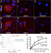
COS-7 cells were transfected with plasmids encoding Drosophila (A,B,C) or human (D,E,F) Dscam. The cells were incubated with medium containing secreted myc epitope tagged Drosophila Netrin B (dNetB; A,D), chick Netrin-1 (cNet-1; B,E), or chick Semaphorin A (cSemaA; C,F). After washing the cells were stained with an anti-myc antibody (red) and counterstained with DAPI (blue). (A) dNetB binding to a cell expressing dDscam. (B) cNet-1 binding to a cell expressing dDscam. (C) cSemaA does not bind to cells expressing dDscam. (D) dNetB binding to a cell expressing hDscamE. (E) cNet-1 binding to cells expressing hDscam. (F) cSemaA does not bind to cells expressing hDscam. (G) Flow cytometry quantitation of the binding of dNetB to COS-7 cells expressing dDscam (blue and green lines). Control cells not expressing dDscam and not incubated with dNetB (red line), and control cells incubated with dNetB (orange). The peaks between the controls and the samples are separate, but the shoulders overlap due to low transfection efficiency of the dDscam construct. (H) The binding of hDscam and rDCC to increasing concentrations of myc tagged Netrin were measured using a HRP conjugated secondary antibody. Scatchard analysis of the data gives dissociation constants of 29.1+/−2.5nM for rDCC/netrin-myc and 35.8+/−8.6nM for hDSCAM/netrin-myc. This difference is not statistically significant (p=0.19, t-test).
Figure 3
Figure 3. Dscams and abl genetically interact in CNS axon guidance
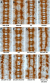
Ventral nerve cords from stage 16 embryos stained with monoclonal antibody BP102 to visualize the CNS axon scaffold with the characteristic pattern of segmentally repeated commissures crossing the CNS midline, and linked by longitudinal tracts. Note the separation of the anterior and posterior commissures by two midline glia, which are separated by a centrally projecting axon (arrow in A). (A) Wild type CNS axon scaffold. (B) Embryos lacking NetA,B (NP5 deficiency) displaying thin and absent commissures (arrowheads), and irregular longitudinal tracts (arrow). (C) fra homozygote showing thin or absent commissures (arrowhead), and altered longitudinal tracts (arrow). (D) Dscam homozygote, which closely resembles wild type. The arrow indicates two midline glia that have not completely separated the commissures, although both are visible. The arrowhead indicates another incomplete separation. The commissures and longitudinal tracts are otherwise unaffected. (E) Dscam3 homozygote. The arrowhead indicates two midline glia that have not fully separated the commissures (subtle). (F) Embryo lacking zygotic abl function. The arrowhead indicates a failure to separate the anterior and posterior commissures. (G) Dscam3 abl homozygote, showing several segments in which the commissures have failed to separate (arrows). (H) Dscam abl homozygote, displaying severe disruption of the commissures. (I) Embryo mutant for Dscam, fra and abl showing an absence of commissures. (J) Dscam Dscam3 double mutant showing disrupted separation of commissures (arrow), and an overall irregularity to the CNS axon scaffold. (K) Dscam fra homozygote displaying greatly reduced or absent commissures, and disrupted longitudinal tracts (arrow). (L) Dscam fra Dscam3 triple mutant homozygote with very few axons crossing the midline, and significantly disrupted longitudinal axon tracts. The residual staining at the midline appears to originate from neurons whose cell bodies reside in the midline (VUMs; arrowheads).
Figure 4
Figure 4. Analysis of identified commissural axons in Dscam mutant combinations
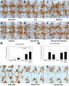
Embryos stained with anti-Connectin antibody (A–F) or the egl-GAL4; UAStaulacZ reporter (G–I). Connectin staining highlights the SP1 cell body (arrows) and commissural axon which crosses the midline in the anterior commissure (arrowheads). All embryos are late stage 15 or early 16, with anterior to the left. (A) Wild type embryo. Note the regular position of the SP1 cell bodies (arrows) and the commissural axons crossing the midline in the anterior commissure (arrowhead). (B) fra mutant showing altered SP1 cell body position (arrow), but intact SP1 axon (arrowhead). The posterior commissure is clearly disrupted in the same segment, as is the adjacent longitudinal. (C) NetA,B mutant showing normal and disrupted SP1 cell body migration (arrows) and missing commissural axons (arrowhead). (D) Dscam mutant embryo, which is nearly identical to wild type, except for some Connectin positive axons inappropriately crossing the midline between the two commissures (asterisk). (E) Dscam fra double mutant displaying missing commissural axons (arrowhead) between the two SP1 cell bodies (arrows; one slightly out of focus). (F) Dscam fra Dscam3 triple mutant showing missing commissural axons between mispositioned SP1 cell bodies (arrows). (G,H) Graphs of SP1 axon midline crossing defects (G) and SP1 cell body migration defects (H) in mutant combinations. Abdominal segments one through seven were scored for ten embryos for each genotype. Statistical significance was assessed by fitting the data to a Poisson distribution. (G) SP1 axon defects. Dscam fra Dscam3 embryos are statistically different from fra mutant embryos (p=0.013), indicating that Dscams enhance the fra SP1 axon phenotype. Dscam fra Dscam3 embryos were statistically different from all other mutants (p<0.01), except Dscam fra. Dscam fra embryos were statistically different from OregonR, NetA,B and Dscam embryos (p<0.05), but were not found to be significantly different from fra alone. (H) Graph of SP1 cell body migration defects. The Dscam and OregonR genotypes are statistically different from all other genotypes except each other (p<0.05). There is no statistical difference between the NetA,B, fra, Dscam fra and Dscam fra Dscam3 phenotypes, indicating that Dscam and Dscam3 do not enhance the fra SP1 cell migration defect. (I) Wild type embryo showing expression of the egl-GAL4;UAS-tau-lacZ transgene. The EW axons cross the midline in the posterior commissure (arrowhead), and the EG cross in the anterior commissure (arrow). (J,K) Two focal planes of one embryo, mutant for Dscam fra. The arrows indicate an EG axon bundle, crossing the midline in an adjacent posterior commissure in response to a missing anterior commissure. The arrowheads indicate EW axons either failing to cross the midline (top) or crossing the midline in an adjacent posterior commissure in response to an absent anterior commissure. The egl-GAL4;UAS-tau-lacZ transgenes do not switch on lacZ in all egl-positive cells in each segment; we have found no evidence of altered cell fate, or missing cells in Dscam mutant combinations.
Figure 5
Figure 5. Dscam misexpression phenotypes
Stage 17 embryos stained with anti-FasII MAb 1D4 (A–E), and stage 16 embryos stained with MAb BP102 (G,H). (A) Wild type embryo in which FasII positive longitudinal fascicles do not cross the midline. (B) Embryo carrying one copy each of the ftzngGAL4 and UAS-Dscam1-30-30-2 transgenes showing a FasII positive fascicle crossing the midline (arrowhead), and also stalled axons (arrow). (C) Embryo mutant for NetA,B and carrying the ftzng-GAL4 transgene, showing a collapse of the longitudinal fascicles into one fascicle (arrow) but no ectopic midline crossing. (D) Embryo mutant for NetA,B and carrying one copy of the ftzng-GAL4 and UAS-Dscam1-30-30-2 transgenes, in which thick (arrowhead) and thin (arrow) fascicles can be seen ectopically crossing the midline. The staining below the thick fascicle is a combination of staining in the transverse nerve and residual staining in cell bodies above and below the plane of focus of the longitudinal axons respectively. (E) Embryo carrying two copies each (based on proportion of embryos displaying this phenotype) of the ftzngGAL4 and UAS-Dscam1-30-30-2 transgenes showing disrupted longitudinal fascicles (arrows) and ectopic axon crossing of the CNS midline (arrowhead). (F) Graph quantifying midline crossing by FasII positive axons in ftzngGAL4 UAS-Dscam1-30-30-2, NetA,B ftzngGAL4 UAS-Dscam1-30-30-2 and NetA,B ftzngGAL4 embryos. There is no statistical difference in the amount of midline crossing observed when NetA,B is deleted (Poisson regression; n=15 for each category). (G) Wild type embryo stained with MAb BP102. (H) Embryo carrying two copies of the ftzngGAL4 and UAS-Dscam1-30-30-2 transgenes showing disrupted separation of the anterior and posterior commissures (arrowhead), and disrupted longitudinal axon tracts (arrow).
Figure 6
Figure 6. DSCAM is expressed by commissural axons in the mouse CNS
Sections of embryos were labeled with antibodies against DSCAM and neuron-specific βIII-tubulin. (A–C) DSCAM is expressed in many neurons in the developing spinal cord. The protein is also detected in commissural axons. (A) Anti-DSCAM antibody labeling of E11.5 spinal cord, including motor column (MN), and commissural axons crossing the floor plate (FP). The dorsal root ganglia (DRG) are labeled as well. (B) The pan-neuronal β-Tubulin (β-IIITub) labeling of the same section shows the location of neurons. (C) Merge of A and B. DSCAM localizes to an inner subset of the commissural fibers (yellow, arrow). (D and E) DSCAM labeling of sections of rhombomere 1 of the E10.5 hindbrain. (D) Both neuron cell bodies and axons are labeled by anti-DSCAM. (E) A close-up of the boxed region in D shows strong DSCAM signal in the hindbrain commissure. Scale Bars: 100 µm in A and 50 µm in D.

References

    1. Agarwala KL, Nakamura S, Tsutumi Y, Yamakawa K. Down syndrome cell adhesion molecule DSCAM mediates homophilic intercellular adhesion. Brain Res. Mol. Brain Res. 2000;79:118–126. - PubMed
    1. Agarwala KL, Ganesh S, Amano K, Suzuki T, Yamakawa K. DSCAM, a highly conserved gene in mammals, expressed in differentiating mouse brain. Biochem. Biophys. Res. Commun. 2001;281:697–705. - PubMed
    1. Barlow GM, Chen XN, Shi ZY, Lyons GE, Kurnit DM, Celle L, Spinner NB, Zackai E, Pettenati MJ, Van Riper AJ, et al. Down syndrome congenital heart disease: a narrowed region and a candidate gene. Genet. Med. 2001;3:91–101. - PubMed
    1. Barlow GM, Micales B, Chen XN, Lyons GE, Korenberg JR. Mammalian DSCAMs: roles in the development of the spinal cord, cortex, and cerebellum? Biochem. Biophys. Res. Commun. 2002;293:881–891. - PubMed
    1. Bashaw GJ, Kidd T, Murray D, Pawson T, Goodman CS. Repulsive axon guidance: Abelson and Enabled play opposing roles downstream of the roundabout receptor. Cell. 2000;101:703–715. - PubMed

Publication types

MeSH terms

LinkOut - more resources