. 2008 Oct 24;322(5901):576-80.
doi: 10.1126/science.1162042.

Midbody targeting of the ESCRT machinery by a noncanonical coiled coil in CEP55

Affiliations

Midbody targeting of the ESCRT machinery by a noncanonical coiled coil in CEP55

Hyung Ho Lee et al. Science. .

Abstract

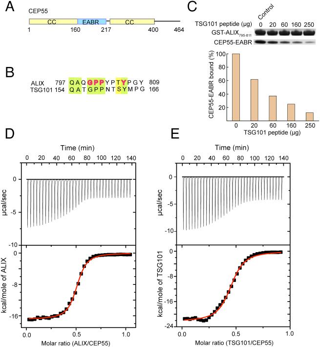
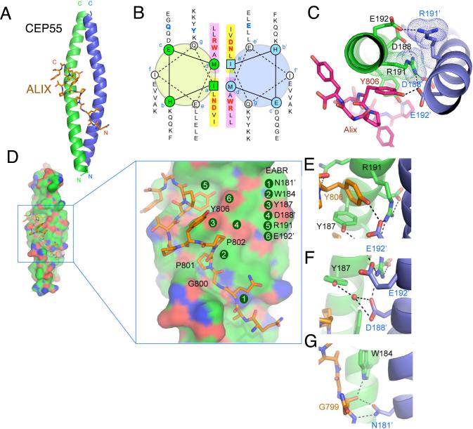
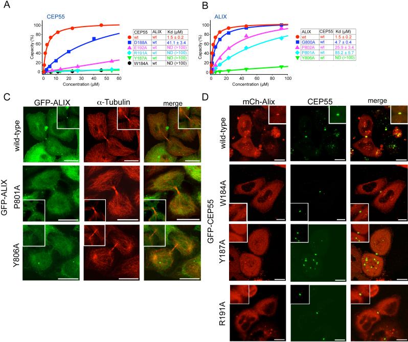
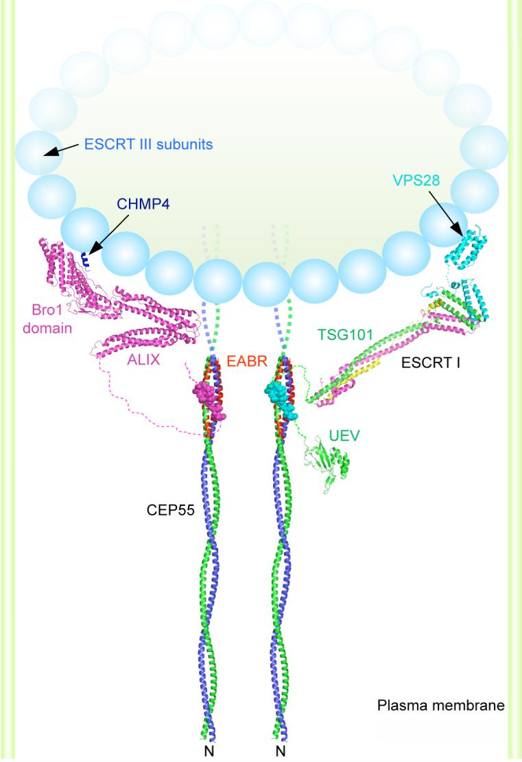
The ESCRT (endosomal sorting complex required for transport) machinery is required for the scission of membrane necks in processes including the budding of HIV-1 and cytokinesis. An essential step in cytokinesis is recruitment of the ESCRT-I complex and the ESCRT-associated protein ALIX to the midbody (the structure that tethers two daughter cells) by the protein CEP55. Biochemical experiments show that peptides from ALIX and the ESCRT-I subunit TSG101 compete for binding to the ESCRT and ALIX-binding region (EABR) of CEP55. We solved the crystal structure of EABR bound to an ALIX peptide at a resolution of 2.0 angstroms. The structure shows that EABR forms an aberrant dimeric parallel coiled coil. Bulky and charged residues at the interface of the two central heptad repeats create asymmetry and a single binding site for an ALIX or TSG101 peptide. Both ALIX and ESCRT-I are required for cytokinesis, which suggests that multiple CEP55 dimers are required for function.


Figures

Figure 1
Figure 1. The CEP55 ESCRT-I and ALIX binding region and its interactions
(A) Predicted coiled-coil regions (yellow) and the EABR of CEP55 (blue). The region designated EABR corresponds to the region formerly suggested to be a hinge between the N- and C-terminal coiled coils (13). (B) CEP55 binding sequences of ALIX and the ESCRT-I subunit TSG101, with conserved residues highlighted with light green shading, and residues shown to be functionally important in ALIX highlighted in bold red type. (C) Pull-down of CEP55-EABR by the GST-ALIX fragment shown, in the presence of the indicated amounts of TSG101 peptide competitor. (D) ITC titration of ALIX peptide into CEP55-EABR solution. (E) ITC titration of TSG101 peptide into CEP55-EABR solution.
Figure 2
Figure 2. Structure of the non-canonical CEP55-EABR coiled coil and its complex with ALIX
(A) The overall structure of the CEP55-EABR homodimer (green and blue ribbons) in complex with the ALIX peptide (stick model, carbon orange, oxygen red, nitrogen blue). At right, the intercoil Cα-Cα distance at the a and d positions of the coiled-coil is shown as a function of residue number for CEP55 (red curve) as compared to the average Cα-Cα distance at the a and d positions of the GCN4 leucine zipper (blueline). (B) Helical wheel analysis of the six heptad repeats of the EABR coiled coil. (C) Charge repulsion between Asp188, Arg191, and Glu192 pairs in the homodimer creates asymmetry in the coiled coil and interactions with the ALIX peptide. (D) Overview and close-ups of selected regions of the CEP55 surface (carbon green, oxygen red, nitrogen blue), with the ALIX peptide colored as in (A). (E-G) Molecular interactions in the complex shown in detail and colored as in (A).
Figure 3
Figure 3. Point mutations in CEP55 and ALIX abrogate binding and cellular localization
(A) SPR analysis of wild-type and mutant CEP55-EABR mutants binding to wild-type ALIX fragment. Binding curves are colored according to affinity in the order red > blue > pink > cyan > green > black in (A) and (B). (B) SPR analysis of wild-type and mutant ALIX fragment binding to wild-type CEP55 EABR. (C) Midbody localization of wild-type and mutant GFP-ALIX expressed in HeLa cells. A magnification of the midbody region is shown in the insets. α−tubulin staining was used (middle panel) to highlight the midbody microtubule structure. (D) Live cell imaging of midbody localization of wild-type mCh-ALIX and wild-type or mutant CEP55 coexpressed in HeLa cells. CEP55 midbody localization was not affected in the mutant (Fig. S7). Insets show higher magnification of the midbody region. Scale bar = 10 μm.
Figure 4
Figure 4. A model for the organization of CEP55-ESCRT and CEP55-ALIX complexes in the midbody
Model of the N-terminal half of the CEP55 structure docked to the ESCRT-I core (24) and UEV domain(21) and ALIX(25) structures as described in the on line methods supplement. The crystallized EABR portion of the CEP55 coiled coil is highlighted in red. The structure of the C-terminal domain of the yeast Vps28 subunit of ESCRT-I is shown (27) as a putative binding site for Vps20, the yeast ortholog of the human ESCRT-III subunit CHMP6. The binding site on the Bro1 domain of ALIX for the C-terminal helix (blue) of the CHMP4 subunit of ESCRT-III is shown (26, 28). A schematic of an ESCRT-III circular array (15) is shown. The width of the membrane neck is not to scale.

References

    1. Glotzer M. Science. 2005;307:1735–1739. - PubMed
    1. Barr FA, Gruneberg U. Cell. 2007;131:847–860. - PubMed
    1. Prekeris R, Gould GW. J. Cell Sci. 2008;121:1569–1576. - PMC - PubMed
    1. Zhao WM, Seki A, Fang GW. Mol. Biol. Cell. 2006;17:3881–3896. - PMC - PubMed
    1. Martinez-Garay I, Rustom A, Gerdes HH, Kutsche K. Genomics. 2006;87:243–253. - PubMed

Publication types

MeSH terms

Associated data