. 2008 Oct 28;105(43):16519-24.
doi: 10.1073/pnas.0804187105. Epub 2008 Oct 23.

Insights into the structural dynamics of the Hsp110-Hsp70 interaction reveal the mechanism for nucleotide exchange activity

Affiliations

Insights into the structural dynamics of the Hsp110-Hsp70 interaction reveal the mechanism for nucleotide exchange activity

Claes Andréasson et al. Proc Natl Acad Sci U S A. .

Abstract

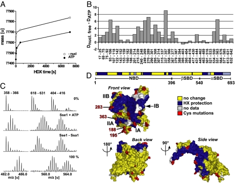
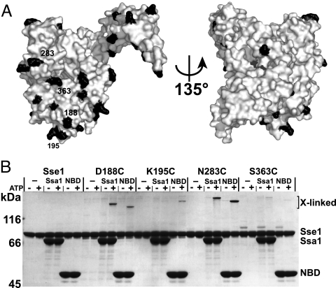
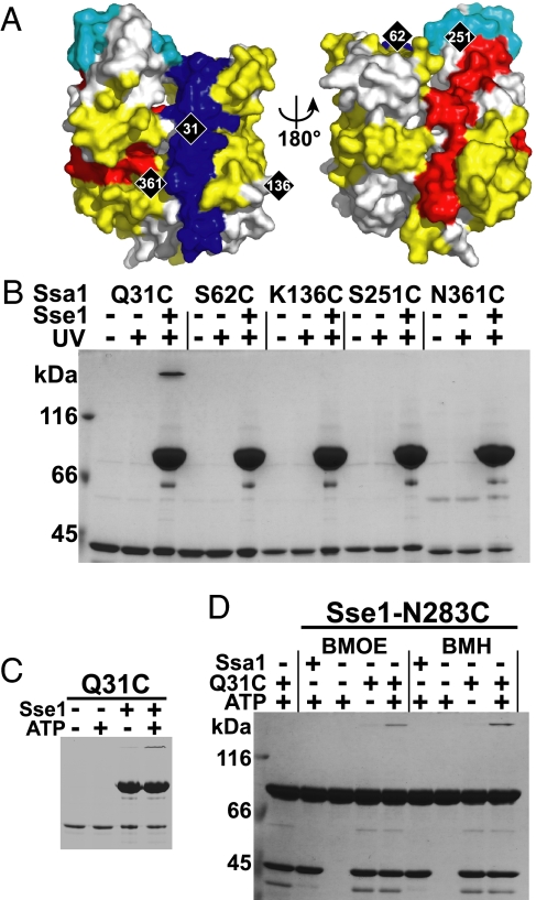
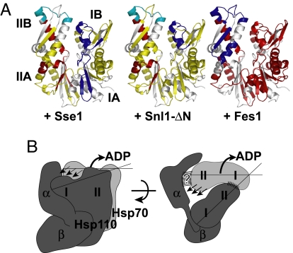
Hsp110 proteins are relatives of canonical Hsp70 chaperones and are expressed abundantly in the eukaryotic cytosol. Recently, it has become clear that Hsp110 proteins are essential nucleotide exchange factors (NEFs) for Hsp70 chaperones. Here, we report the architecture of the complex between the yeast Hsp110, Sse1, and its cognate Hsp70 partner, Ssa1, as revealed by hydrogen-deuterium exchange analysis and site-specific cross-linking. The two nucleotide-binding domains (NBDs) of Sse1 and Ssa1 are positioned to face each other and form extensive contacts between opposite lobes of their NBDs. A second contact with the periphery of the Ssa1 NBD lobe II is likely mediated via the protruding C-terminal alpha-helical subdomain of Sse1. To address the mechanism of catalyzed nucleotide exchange, we have compared the hydrogen exchange characteristics of the Ssa1 NBD in complex with either Sse1 or the yeast homologs of the NEFs HspBP1 and Bag-1. We find that Sse1 exploits a Bag-1-like mechanism to catalyze nucleotide release, which involves opening of the Ssa1 NBD by tilting lobe II. Thus, Hsp110 proteins use a unique binding mode to catalyze nucleotide release from Hsp70s by a functionally convergent mechanism.


Conflict of interest statement

The authors declare no conflict of interest.

Figures

Fig. 1.
Fig. 1.
Ssa1 association induces HX protection in both the front face of the NBD and the C-terminal α-helical domain of ATP-bound Sse1. (A) Stabilization effect of ATP binding to Sse1, as monitored from the amide proton exchange properties of the protein. Preincubated protein samples in the presence (filled symbols) or absence (open symbols) of ATP were diluted 20-fold into D2O buffer and incubated for different time intervals at 30 °C before quenching the proton–deuteron exchange reaction. The mass increase through deuteron incorporation was determined for Sse1. (B) Difference in deuteron incorporation between nucleotide-free Sse1 and Sse1 + ATP. The data were resolved to individual peptic peptides as indicated by the start and end residue numbers of the corresponding segments (Table S1). (C) HX properties of Sse1 + ATP and Sse1 bound to Ssa1. Mass spectra of representative peptides for protein samples incubated in H2O (0%) or for 2 min in D2O buffer. 100% shows a control spectrum of the same peptide from fully deuterated Sse1. (D) Observed HX protection pattern induced by Ssa1 association, derived from comparison of Sse1 + ATP and Sse1–Ssa1 (see C and Fig. S2) (13). The color code for the peptides is as follows: blue, Ssa1-induced protection; yellow, no change in HX properties upon association with Ssa1; and gray, no data available. The 29 residues that were mutated to cysteine for cross-linking experiments are marked in red (residue numbers 22, 34, 98, 135, 188, 194, 195, 218, 222, 254, 283, 290, 304, 331, 346, 355, 363, 374, 450, 529, 566, 576, 586, 595, 609, 630, 639, 650, and 654). The first projection corresponds to the standard view of Hsp70 NBD, and the domain nomenclature follows Flaherty et al. (23).
Fig. 2.
Fig. 2.
Positions at Sse1 NBD subdomain IIA and IIB outer rim cross-link to Ssa1. Each mutant protein (see Fig. 1D) was BPIA-labeled on its introduced cysteine and tested for the ability to cross-link to full-length or the NBD of Ssa1 as described in Materials and Methods. Four BPIA-labeled cysteines formed ATP-dependent cross-links to Ssa1.
Fig. 3.
Fig. 3.
The α-helical SBD subdomain is important for the interaction with Ssa1. (A) Schematic representation of the truncation mutants of Sse1. The α-helical subdomain (α) is drawn as a ribbon structure (13) with names of helices and the last residue of each truncation mutant indicated. (B) Ten-fold serial dilutions of sse1Δ sse2Δ or sse1Δ sse2Δ fes1Δ (Δ) yeast strains carrying SSE1, or derived truncated alleles, in the context of its endogenous locus on a single-copy plasmid were spotted onto YPD medium and incubated at 30 °C or 37 °C for 2 days. (C) MABA-ADP dissociation rate [koff (s−1)] from Ssa1 (0.5 μM) by the indicated concentrations of Sse1 or derived truncation mutants. The Sse1 proteins were preincubated with 3 mM ATP before measuring the release rates with a stopped-flow instrument.
Fig. 4.
Fig. 4.
Residue 31 on the front face of Ssa1 cross-links with residue 283 on the front face of Sse1. (A) Ssa1 NBD modeled onto the structure of the Hsc70 NBD (23) as viewed from the front and back. The molecule is colored according to the observed pattern of Sse1-induced HX protection (blue), weak protection (cyan), and deprotection (red) (Fig. S5). Yellow indicates no change in HX properties, and gray, no data available. Positions of the 5 introduced cysteines are marked in black. (B) Each mutant protein was BPIA-labeled on its introduced cysteine and tested for the ability to cross-link to Sse1 (Upper). (C) The cross-link at Q31C was tested for ATP dependence (Lower). (D) Sse1-N283C was mixed with the control protein Ssa1 NBD (Ssa1) or the Ssa1 NBD carrying the Q31C mutation (Q31C) in the presence or absence of 1 mM ATP and tested for its ability to form a cysteine–cysteine cross-link with BMOE and BMH.
Fig. 5.
Fig. 5.
Sse1 utilizes a Bag-1-type NEF mechanism. (A) Pattern of NEF-induced HX protection and deprotection mapped onto a model of Ssa1-NBD. The effects of Sse1 (Left), Snl1-ΔN (Center), and Fes1 (Right) were compared in 10-s HX reactions (Fig. S8). For the color code, see Fig. 4A. (B) Schematic model for Hsp110 (dark gray) and Hsp70 (light gray) interaction and the mechanism of catalyzed nucleotide release. The lobes (I/II) of the 2 respective NBDs and the subdomains of the Hsp110 SBD (α/β) are indicated. Striped areas mark interaction surfaces. Arrows denote outward rotation of Hsp70 NBD lobe II and the triggered release of ADP. See Discussion for details.

References

    1. Craig EA, Huang P. In: Protein Folding Handbook. Buchner J, Kiefhaber T, editors. Wiley; 2005. pp. 490–515.
    1. Mayer MP, Bukau B. Hsp70 chaperones: Cellular functions and molecular mechanism. Cell Mol Life Sci. 2005;62:670–684. - PMC - PubMed
    1. Harrison CJ, Hayer-Hartl M, Di Liberto M, Hartl F, Kuriyan J. Crystal structure of the nucleotide exchange factor GrpE bound to the ATPase domain of the molecular chaperone DnaK. Science. 1997;276:431–435. - PubMed
    1. Sondermann H, et al. Structure of a Bag/Hsc70 complex: Convergent functional evolution of Hsp70 nucleotide exchange factors. Science. 2001;291:1553–1557. - PubMed
    1. Shomura Y, et al. Regulation of Hsp70 function by HspBP1: Structural analysis reveals an alternate mechanism for Hsp70 nucleotide exchange. Mol Cell. 2005;17:367–379. - PubMed

Publication types

MeSH terms

LinkOut - more resources