. 2008 Nov 21;103(11):1280-8.
doi: 10.1161/CIRCRESAHA.108.183863. Epub 2008 Oct 23.

Mutation in EGFP domain of LDL receptor-related protein 6 impairs cellular LDL clearance

Affiliations

Mutation in EGFP domain of LDL receptor-related protein 6 impairs cellular LDL clearance

Wenzhong Liu et al. Circ Res. .

Abstract

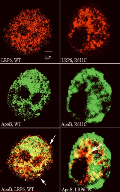
Mutation in the EGFP domain of LDL receptor-related protein 6 (LRP6(R611C)) is associated with hypercholesterolemia and early-onset atherosclerosis, but the mechanism by which it causes disease is not known. Cholesterol uptake was examined in cells from LRP6(+/-) mice and LRP6(R611C) mutation carriers. Splenic B cells of LRP6(+/-) mice have significantly lower LRP6 expression and low-density lipoprotein (LDL) uptake than those of the wild-type littermates. Although similar levels of total LRP6 were found in lymphoblastoid cells (LCLs) of LRP6(R611C) mutation carriers and those of the unaffected family member, LDL uptake was significantly lower in the mutant cells. Mutant and wild-type receptors show similar affinities for apolipoprotein B at neutral pH. LRP6 colocalized with LDL and was coimmunoprecipitated with NPC1 (Niemann-Pick disease type C1), an endocytic regulator of LDL trafficking. However, the cellular localization of LRP6 in the mutant cells shifted from cell surface to late endosomes/lysosomes. Plasma membrane expression levels of LRP6(R611C) was lower compared to wild-type receptor and declined to a greater extent in LDL-rich medium. Further examinations revealed lower efficacy of apolipoprotein B dissociation from LRP6(R611C) compared to wild-type receptor at an acidic pH. These studies identify LRP6 as a receptor for LDL endocytosis and imply that R611C mutation results in reduced LRP6 membrane expression and decreased LDL clearance. Based on our findings, we conclude that the increased affinity of the mutant receptor for LDL in acidic pH leads to their impaired dissociation in late endosomes, which compromises their recycling to the plasma membrane.


Figures

Figure 1
Figure 1. Greater LDL uptake of NIH3T3 cells transfected with wildtype LRP6 compared to those of untransfected cells and cells transfected with LRP6R611C (1a), reduced LDL uptake of splenic B lymphocytes of the LRP6+/ mice compared to those of the wildtype littermates (1b) and increased LDL uptake of wildtype lymphocytes compared to LRP6R611C lymphocytes (1c) despite equal membrane expression of the LDL receptor (1d), total LRP6, LDLR and SREBP (1e, 1f)
1a- NIH3T3 cells were transfected with plasmids encoding hemagglutinin-tagged wild-type (WT), or mutant LRP6 or with empty plasmids. Cells were incubated with Dil-LDL at 37°C and subsequently fixed in 4 % paraformaldehyde. Single cell fluorescence intensities were measured using fluorescence activated cell sorter (FACS). 1b- LDL uptake of splenic B lymphocytes of the LRP6+/ mice compared to those of the wildtype littermates was explored using same technique. LRP6R611C cells demonstrate lower LDL uptake (29 ±3.4 %, p<0.005) compared to wildtype cells after 1 hr incubation with Dil-LDL. Lower panel shows expression levels of LRP6 in WT and LRP6+/ lymphocytes. 1c- LDL uptake of immortalized lymphoblastoid cells (LCL) of the LRP6R611C mutation carriers (R611C) and their unaffected family members (WT) was explored using same technique. LRP6R611C cells demonstrate lower LDL uptake (31 ±2.7 %, p<0.005) compared to wildtype cells after 4 hr incubation with Dil-LDL. 1d- Plasma membrane LDLR expression, assayed by fluorescence activated cell sorter (FACS), is similar in LCLs of the LRP6R611C mutation carriers and their unaffected family members. 1e- mRNA and 1f- protein expression of LRP6, LDLR and SREBP1 (Sterol regulatory binding protein 1) are roughly equal in the mutant and wildtype cells, while total LDLR mRNA expression in slightly higher in the mutant cells.
Fig 2
Fig 2. LRP6 colocalizes with LDL and its cellular distribution is perturbed in the mutant cells
(2a) LDL colocalizes with LRP6 and is abnormally redistributed in the mutant cells. The upper panel demonstrates LRP6 localization (Cy2) in the wildtype (WT) and mutant LCLs (R611C). Goat anti LRP6 was used as the primary antibody. The middle panel shows Dil-LDL uptake of the same cells. Dil-LDL colocalizes with LRP6 in the wildtype and mutant cells (lower panel). The Colocalization is shifted away from cell surface seen in the wildtype cells to clusters within the cell in the mutant cells. Arrow head shows clustering of LDL and arrows show Colocalization of Dil-LDL with LRP6. (2b) LRP6 is localized evenly along the endocytotic pathway as seen by Alexa Fluor® 488-transferrin in the wildtype cells (left panels) but is shifted away from the cell surface to the late endocytotic compartments in the mutant cells (right panels). (2c) similarly while lesser amount of the mutant LRP6 localizes to early endosomes (right panels) compared to wildtype LRP6 (2d) significantly higher amount of the mutant LRP6 is localized to late endosomes/lysosomes labeled with LAMP2b (right panel) compared to the wildtype LRP6 (left panel).
Fig. 3
Fig. 3
a- ApoB binds wildtype and mutant LRP6. The affinity of the mutant and wildtype LRP6 for apoB in neutral pH is demonstrated. BSA was used as control. Signals were detected by assaying binding of apoB antibody by ELISA. The results are shown as mean and standard errors of quadruplicate experiments. The affinity of the mutant LRP6 for apoB was slightly higher compared to wildtype receptor. OD, optic density, BSA, bovine serum albumin. Fig. 3b- ApoB colocalizes with LRP6 in wildtype cells at the cell surface and within the cell (arrows, lower left panel). In contrast mutant LRP6-apoB Colocalization is less in the cell periphery and more in the juxtanuclear region (right panels). The upper panel demonstrates LRP6 localization (Cy2) in the wildtype (WT) and the mutant LCLs (b). The middle and lower panels show cellular localization of apoB and its colocalization with LRP6 respectively (see arrows).
Fig. 3
Fig. 3
a- ApoB binds wildtype and mutant LRP6. The affinity of the mutant and wildtype LRP6 for apoB in neutral pH is demonstrated. BSA was used as control. Signals were detected by assaying binding of apoB antibody by ELISA. The results are shown as mean and standard errors of quadruplicate experiments. The affinity of the mutant LRP6 for apoB was slightly higher compared to wildtype receptor. OD, optic density, BSA, bovine serum albumin. Fig. 3b- ApoB colocalizes with LRP6 in wildtype cells at the cell surface and within the cell (arrows, lower left panel). In contrast mutant LRP6-apoB Colocalization is less in the cell periphery and more in the juxtanuclear region (right panels). The upper panel demonstrates LRP6 localization (Cy2) in the wildtype (WT) and the mutant LCLs (b). The middle and lower panels show cellular localization of apoB and its colocalization with LRP6 respectively (see arrows).
Fig. 4
Fig. 4. Cell surface expression of the mutant LRP6 is reduced
Compared to wildtype LCLs, lymphoblastoid cells of the LRP6R611C mutation carriers demonstrate lower membrane LRP6 expressions in LDPDS assayed by flow cytometry. Rabbit anti-LRP6 was used as the primary antibody (4a). Expression levels of the membrane LRP6 falls more drastically in the mutant LCLs compared to wildtype LCLs in LDL-enriched medium. This effect is reversible in the wildtype cells. Roughly after 4 hours incubation in LPDS, the wildtype receptor expression approaches its normal level, but the mutant receptor expression is modestly increased (4b). The results are shown as mean of quadruplicate experiments in lymphoblastoid cell lines of four mutation carriers and four unaffected family members.
Figure 5
Figure 5. Binding and colocalization of LRP6 with NPC1 confirms its involvement in endosomal LDL trafficking
5a- The immunecoprecipitation of LRP6 with NPC1 is demonstrated. Goat anti-LRP6 and rabbit anti-NPC-1 polyclonal antibodies were used. Lane 1: wild type (left) and mutant LRP6 (right). Lane 2: Wild type NPC1 (left) and mutant NPC1 (right). The absence of binding to NPC-2 and PCSK9 are shown in lanes 4 and 6 respectively 5(b) LRP6 and NPC1 colocalize in mutant and wildtype cells. The upper panel demonstrates LRP6 staining (Cy-2) and the middle panel shows the NPC1 in the wildtype (WT) and mutant cells (R611C). The lower panel shows colocalizations of LRP6 and NPC1 (see arrows). Colocalization in the wildtype cells is mainly in the periphery and in the mutant cells in the juxtanuclear regions.
Fig. 6
Fig. 6. The binding affinity of plasma apoB to LRP6 is greater in the mutant than WT in acidic pH
The affinity of the mutant and wildtype LRP6 for apoB in acidic pH is demonstrated. The results are shown as mean and standard errors of quadruplicate experiments. BSA was used as control. The affinity of the mutant LRP6 for apoB was significantly higher compared to wildtype receptor. OD, optic density, BSA, bovine serum albumin.

Similar articles

Cited by

References

    1. Llorente-Cortes V, Martinez-Gonzalez J, Badimon L. LDL receptor-related protein mediates uptake of aggregated LDL in human vascular smooth muscle cells. Arterioscler Thromb Vasc Biol. 2000;20:1572–9. - PubMed
    1. Magoori K, Kang MJ, Ito MR, Kakuuchi H, Ioka RX, Kamataki A, Kim DH, Asaba H, Iwasaki S, Takei YA, Sasaki M, Usui S, Okazaki M, Takahashi S, Ono M, Nose M, Sakai J, Fujino T, Yamamoto TT. Severe hypercholesterolemia, impaired fat tolerance, and advanced atherosclerosis in mice lacking both low density lipoprotein receptor-related protein 5 and apolipoprotein E. J Biol Chem. 2003;278:11331–6. - PubMed
    1. Fujino T, Asaba H, Kang MJ, Ikeda Y, Sone H, Takada S, Kim DH, Ioka RX, Ono M, Tomoyori H, Okubo M, Murase T, Kamataki A, Yamamoto J, Magoori K, Takahashi S, Miyamoto Y, Oishi H, Nose M, Okazaki M, Usui S, Imaizumi K, Yanagisawa M, Sakai J, Yamamoto TT. Low-density lipoprotein receptor-related protein 5 (LRP5) is essential for normal cholesterol metabolism and glucose-induced insulin secretion. Proc Natl Acad Sci U S A. 2003;100:229–34. - PMC - PubMed
    1. Beisiegel U, Weber W, Ihrke G, Herz J, Stanley KK. The LDL-receptor-related protein, LRP, is an apolipoprotein E-binding protein. Nature. 1989;341:162–4. - PubMed
    1. Kowal RC, Herz J, Goldstein JL, Esser V, Brown MS. Low density lipoprotein receptor-related protein mediates uptake of cholesteryl esters derived from apoprotein E-enriched lipoproteins. Proc Natl Acad Sci U S A. 1989;86:5810–4. - PMC - PubMed

Publication types

MeSH terms

Substances