. 2008 Nov 21;103(11):1270-9.
doi: 10.1161/CIRCRESAHA.108.182832. Epub 2008 Oct 23.

Heat shock protein 20 interacting with phosphorylated Akt reduces doxorubicin-triggered oxidative stress and cardiotoxicity

Affiliations

Heat shock protein 20 interacting with phosphorylated Akt reduces doxorubicin-triggered oxidative stress and cardiotoxicity

Guo-Chang Fan et al. Circ Res. .

Abstract

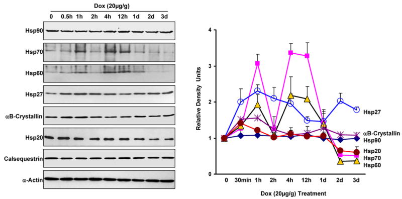
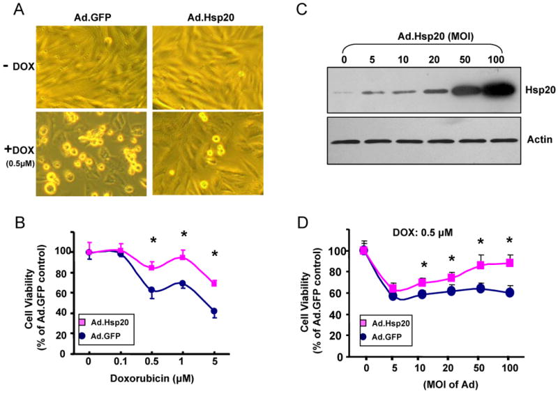
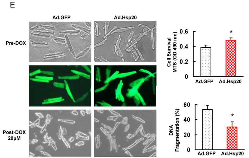
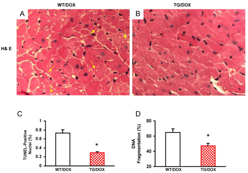
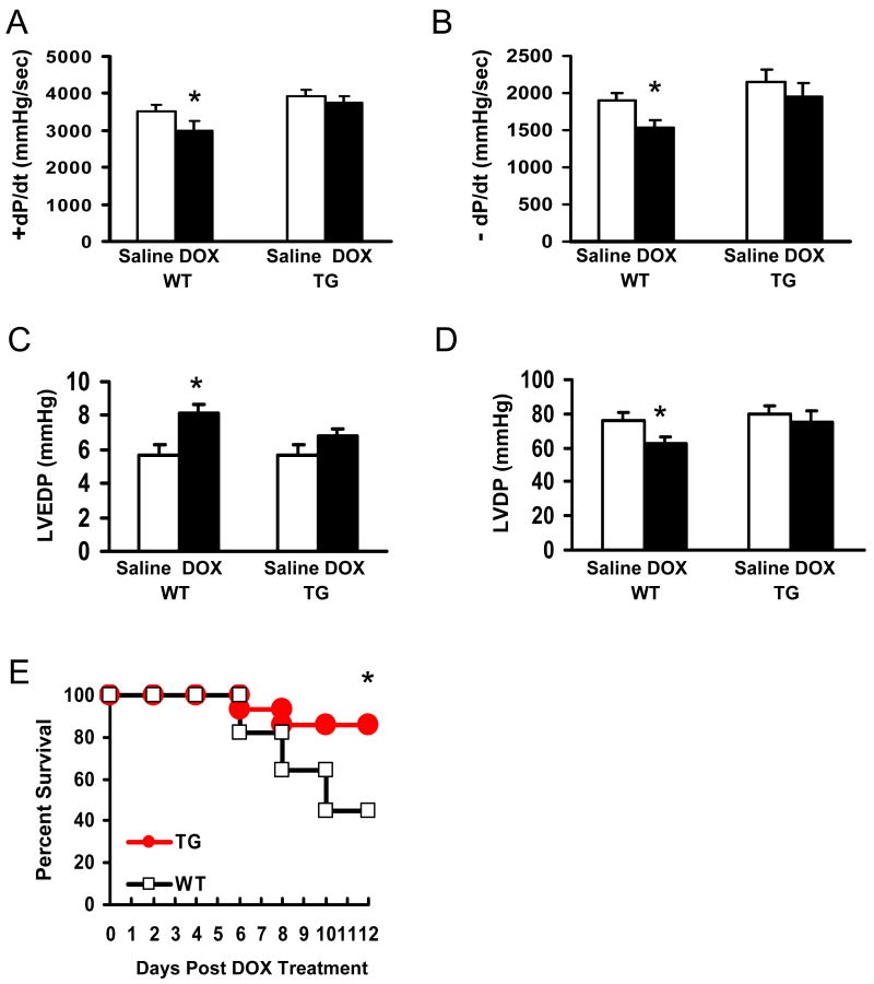
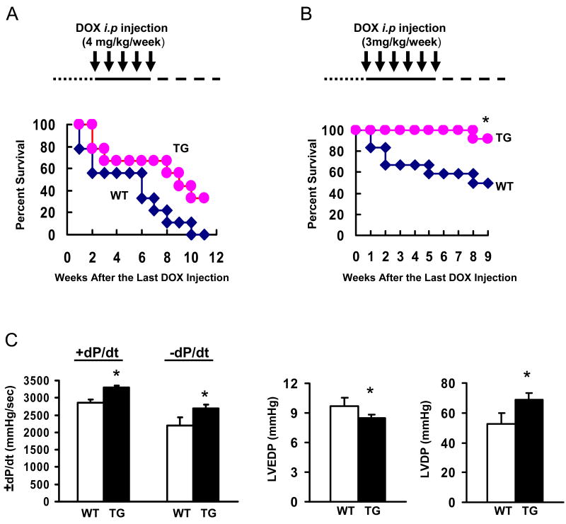
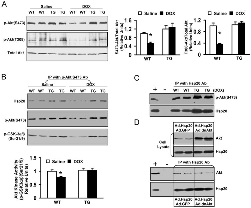
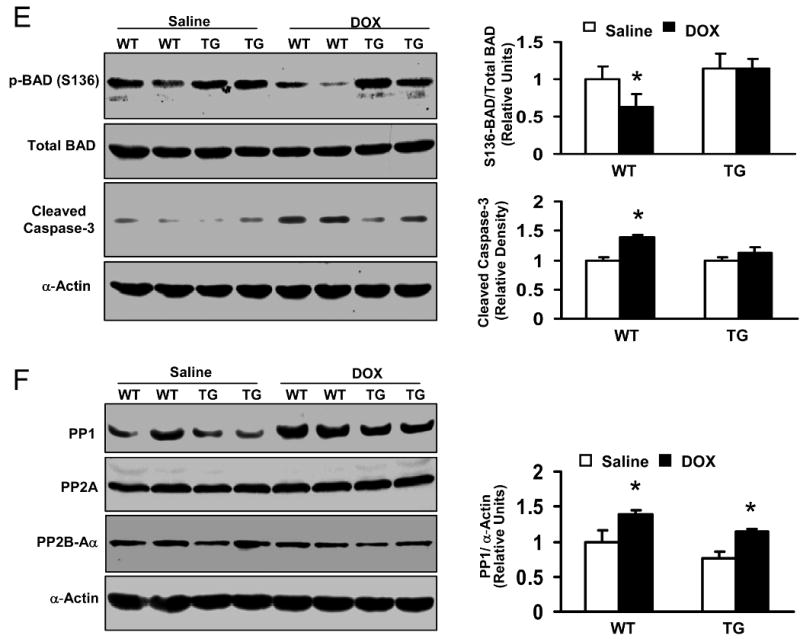
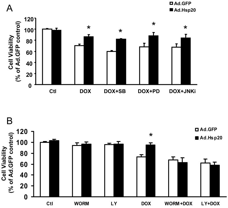
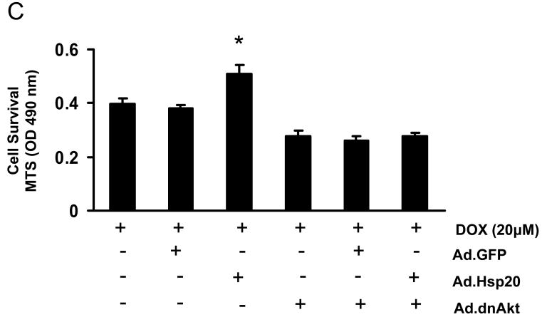
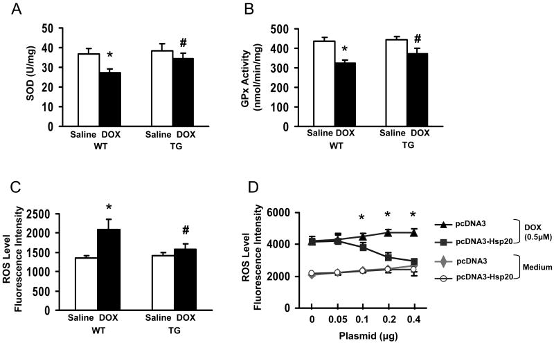
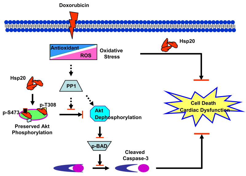
Doxorubicin (DOX) is a widely used antitumor drug, but its application is limited because of its cardiotoxic side effects. Heat shock protein (Hsp)20 has been recently shown to protect cardiomyocytes against apoptosis, induced by ischemia/reperfusion injury or by prolonged beta-agonist stimulation. However, it is not clear whether Hsp20 would exert similar protective effects against DOX-induced cardiac injury. Actually, DOX treatment was associated with downregulation of Hsp20 in the heart. To elucidate the role of Hsp20 in DOX-triggered cardiac toxicity, Hsp20 was first overexpressed ex vivo by adenovirus-mediated gene delivery. Increased Hsp20 levels conferred higher resistance to DOX-induced cell death, compared to green fluorescent protein control. Furthermore, cardiac-specific overexpression of Hsp20 in vivo significantly ameliorated acute DOX-triggered cardiomyocyte apoptosis and animal mortality. Hsp20 transgenic mice also showed improved cardiac function and prolonged survival after chronic administration of DOX. The mechanisms underlying these beneficial effects were associated with preserved Akt phosphorylation/activity and attenuation of DOX-induced oxidative stress. Coimmunoprecipitation studies revealed an interaction between Hsp20 and phosphorylated Akt. Accordingly, BAD phosphorylation was preserved, and cleaved caspase-3 was decreased in DOX-treated Hsp20 transgenic hearts, consistent with the antiapoptotic effects of Hsp20. Parallel ex vivo experiments showed that either infection with a dominant-negative Akt adenovirus or preincubation of cardiomyocytes with the phosphatidylinositol 3-kinase inhibitors significantly attenuated the protective effects of Hsp20. Taken together, our findings indicate that overexpression of Hsp20 inhibits DOX-triggered cardiac injury, and these beneficial effects appear to be dependent on Akt activation. Thus, Hsp20 may constitute a new therapeutic target in ameliorating the cardiotoxic effects of DOX treatment in cancer patients.


Figures

Figure 1
Figure 1
Time course of major Hsps' expression in the mouse heart after i.p. administration of doxorubicin (20 mg/kg). At the indicated intervals, mouse hearts were excised and homogenized to assess major Hsps' expression by Western blot analysis (Left panel). The right panel shows the quantitative results from the Western-blots. Calsequestrin and α-Actin as internal controls (n=4 hearts for each time point).
Figure 2
Figure 2
Effect of increased Hsp20 expression on cardiaomyocyte survival after doxorubicin treatment. (A) Photomicrographs of Ad.Hsp20-infected or Ad.GFP-infected H9c2 cells (50 MOI) were taken in the presence or absence of doxorubicin (0.5 μM) for 24 h. (B) Ad.Hsp20-infected H9c2 cells showed significantly higher cell viability than Ad.GFP-infected cells after treatment with doxorubicin at 0.5, 1 or 5 μM for 24 h. (C) Western-blots showed increased Hsp20 expression upon increased multiplicity of infection (MOI) of adenoviral vector. (D) Protection of H9c2 cells against doxorubicin was shown to be dependent on the Hsp20 expression levels. (E) There was no morphological change between the empty vector Ad.GFP- and Ad.Hsp20-infected myocytes, and more than 95% adult rat cardiomyocytes were infected by recombinant adenovirus vector after 24 h, as indicated by GFP fluorescence (Magnification 200×). Hsp20-overexpressed cardiomyocytes were resistant to doxorubicin-induced cell death, as evidenced by decreased number of round-shaped cardiomyocytes (Magnification 100×), and MTS analysis, as well as DNA fragmentation assay, determined by cell-death-detection ELISA kit (*P < 0.05 versus control Ad.GFP). Similar results were observed in three additional, independent experiments.
Figure 2
Figure 2
Effect of increased Hsp20 expression on cardiaomyocyte survival after doxorubicin treatment. (A) Photomicrographs of Ad.Hsp20-infected or Ad.GFP-infected H9c2 cells (50 MOI) were taken in the presence or absence of doxorubicin (0.5 μM) for 24 h. (B) Ad.Hsp20-infected H9c2 cells showed significantly higher cell viability than Ad.GFP-infected cells after treatment with doxorubicin at 0.5, 1 or 5 μM for 24 h. (C) Western-blots showed increased Hsp20 expression upon increased multiplicity of infection (MOI) of adenoviral vector. (D) Protection of H9c2 cells against doxorubicin was shown to be dependent on the Hsp20 expression levels. (E) There was no morphological change between the empty vector Ad.GFP- and Ad.Hsp20-infected myocytes, and more than 95% adult rat cardiomyocytes were infected by recombinant adenovirus vector after 24 h, as indicated by GFP fluorescence (Magnification 200×). Hsp20-overexpressed cardiomyocytes were resistant to doxorubicin-induced cell death, as evidenced by decreased number of round-shaped cardiomyocytes (Magnification 100×), and MTS analysis, as well as DNA fragmentation assay, determined by cell-death-detection ELISA kit (*P < 0.05 versus control Ad.GFP). Similar results were observed in three additional, independent experiments.
Figure 3
Figure 3
Cardiac-specific overexpression of Hsp20 attenuates cardiomyocyte death and apoptosis after acute administration of DOX. Hematoxylin and eosin (H& E) staining of mouse ventricle [A: wild-type (WT); B: Hsp20 transgenic (TG)] on day 4 after DOX treatment. Arrows indicate representative vacuolization. Quantitative results of TUNEL staining are shown in (C). n=5, with 2 sections from each heart. *P<0.01, compared with DOX-treated WT hearts. (D) DNA fragmentation assay, 200 μg heart homogenate was determined by cell-death-detection ELISA kit (*P < 0.05 versus DOX-treated WT hearts, n=6).
Figure 4
Figure 4
Overexpression of Hsp20 in vivo improves cardiac function after acute administration of DOX (20 mg/kg), as determined by the rates of contraction (+dP/dt) and relaxation (−dP/dt) (A and B), the left ventricular end-diastolic pressure (LVEDP) (C) and the left ventricular developed pressure (LVDP) (D). Hsp20 overexpression also prolongs the animal survival after acute treatment of DOX (E). (*P<0.05; compared with vehicle or WT group, n=5 for function measurement, and n=11-14 for animal survival observation).
Figure 5
Figure 5
Overexpression of Hsp20 in vivo prolongs the animal survival (A: 4mg/kg/week for 5 weeks; B, 3 mg/kg/week for 6 weeks) and improves cardiac function (C) after chronic administration of DOX. Cardiac function was measured by ex vivo Langendorff preparations at 1 week after last i.p injection of DOX (3 mg/kg weekly for 6 times) (*P<0.05; compared with WT group, n=9-12 for animal survival observation, and n=4-5 for functional measurement).
Figure 6
Figure 6
Effects of Hsp20 on the expression of Akt-BAD-caspase3 signaling cascades in the DOX-treated hearts. Hearts were excised on day 4 after acute administration of DOX (20 mg/kg), homogenized with lysis buffer, and subjected to Western-blotting analysis. (A) The phosphorylation levels of Akt at either S473 or T308 site were significantly decreased in WT hearts after DOX treatment, while there were no alterations in Hsp20-TG hearts. (B) In vitro Akt kinase activity assay by using GST-fused GSK-3α/β crosstide corresponding to residues surrounding GSK-3α/β (Ser21/9) as a substrate. This immunoplex was also probed with Ser473-Akt or Hsp20 antibody. The same amount of heart homogenate (1mg/200μl) was immunoprecipited with Ser473-Akt antibody. (C) Reciprocal co-immunoprecipitation with Hsp20 antibody revealed an increased association of Hsp20 with Ser473-Akt in DOX-treated TG hearts. Preimmunoprecipitated WT heart homogenate was used as positive control (+), and immunoprecipitate without Hsp20 antibody (added only protein G PLUS agarose) was used as negative control (-). (D) H9c2 cells were co-infected with a non-phosphorylatable Akt (Ad.dnAkt) and Ad.Hsp20, and 48 hours later, cells were collected and added NP40 lysis buffer. The same amount of cell lysates was subjected to SDS-PAGE or immunoprecipitated with Hsp20 antibody. Preimmunoprecipitated H9c2 cell lysate was used as positive control (+), and immunoprecipitate without Hsp20 antibody was used as negative control (-). (E) 150 μg heart homogenate was immuno-blotted with p-BAD (S136) or BAD antibody, or the cleaved caspase-3 antibody. (F) 100 μg heart homogenate was immuno-blotted with PP1, PP2A, or PP2B-Aα antibody. (n=4 hearts for each group, *P<0.01, compared with saline controls).
Figure 6
Figure 6
Effects of Hsp20 on the expression of Akt-BAD-caspase3 signaling cascades in the DOX-treated hearts. Hearts were excised on day 4 after acute administration of DOX (20 mg/kg), homogenized with lysis buffer, and subjected to Western-blotting analysis. (A) The phosphorylation levels of Akt at either S473 or T308 site were significantly decreased in WT hearts after DOX treatment, while there were no alterations in Hsp20-TG hearts. (B) In vitro Akt kinase activity assay by using GST-fused GSK-3α/β crosstide corresponding to residues surrounding GSK-3α/β (Ser21/9) as a substrate. This immunoplex was also probed with Ser473-Akt or Hsp20 antibody. The same amount of heart homogenate (1mg/200μl) was immunoprecipited with Ser473-Akt antibody. (C) Reciprocal co-immunoprecipitation with Hsp20 antibody revealed an increased association of Hsp20 with Ser473-Akt in DOX-treated TG hearts. Preimmunoprecipitated WT heart homogenate was used as positive control (+), and immunoprecipitate without Hsp20 antibody (added only protein G PLUS agarose) was used as negative control (-). (D) H9c2 cells were co-infected with a non-phosphorylatable Akt (Ad.dnAkt) and Ad.Hsp20, and 48 hours later, cells were collected and added NP40 lysis buffer. The same amount of cell lysates was subjected to SDS-PAGE or immunoprecipitated with Hsp20 antibody. Preimmunoprecipitated H9c2 cell lysate was used as positive control (+), and immunoprecipitate without Hsp20 antibody was used as negative control (-). (E) 150 μg heart homogenate was immuno-blotted with p-BAD (S136) or BAD antibody, or the cleaved caspase-3 antibody. (F) 100 μg heart homogenate was immuno-blotted with PP1, PP2A, or PP2B-Aα antibody. (n=4 hearts for each group, *P<0.01, compared with saline controls).
Figure 7
Figure 7
Cardioprotection of Hsp20 against DOX -induced cell death is dependent on Akt signaling. (A) Pre-treated cardiomyocytes with SB203580, PD98059, or JNK inhibitor had no effect on protection of Hsp20 against DOX-induced cell death. Cardiomyocytes were infected with Ad.GFP or Ad.Hsp20 for 24 h; subsequently, SB203580, PD98059, or JNK inhibitor was added for one hour, and then these cells were incubated with DOX (20 μM) for 24 h. Cell survival was measured by MTS assay. (B) Pre-treatment of the Ad.Hsp20-infected cardiomyocytes with either Ly294002 or wortmannin totally inhibited the protective effects of Hsp20 against DOX. (C) Infection with dominant-negative Akt (Ad.dnAkt) eliminated the protection provided by overexpressed Hsp20 in cardiomyocytes against DOX. *P<0.05; compared with Ad.GFP-infected group. Data shown are representative of 3 separated experiments.
Figure 7
Figure 7
Cardioprotection of Hsp20 against DOX -induced cell death is dependent on Akt signaling. (A) Pre-treated cardiomyocytes with SB203580, PD98059, or JNK inhibitor had no effect on protection of Hsp20 against DOX-induced cell death. Cardiomyocytes were infected with Ad.GFP or Ad.Hsp20 for 24 h; subsequently, SB203580, PD98059, or JNK inhibitor was added for one hour, and then these cells were incubated with DOX (20 μM) for 24 h. Cell survival was measured by MTS assay. (B) Pre-treatment of the Ad.Hsp20-infected cardiomyocytes with either Ly294002 or wortmannin totally inhibited the protective effects of Hsp20 against DOX. (C) Infection with dominant-negative Akt (Ad.dnAkt) eliminated the protection provided by overexpressed Hsp20 in cardiomyocytes against DOX. *P<0.05; compared with Ad.GFP-infected group. Data shown are representative of 3 separated experiments.
Figure 8
Figure 8. Hsp20 overexpression attenuates DOX-triggered oxidative stress
A: superoxide dismutase (SOD); B: glutathione peroxidase (GPx); and (C) amount of ROS, determined by DCF fluorescence intensity. The specificity of DCF signal was confirmed by the use of SOD (Online Figure IV). (n=5 hearts, *P<0.01, # P<0.05, compared to saline controls, respectively). (D) The amount of ROS was decreased in pcDNA3-Hsp20-transfected H9c2 cells in a dose-dependent fashion. *P<0.05; compared with pcDNA3-cells. Data shown are representative of 3 separated experiments. (E) Proposed scheme for protection of Hsp20 against DOX-triggered cardiotoxicity. Treatment of cardiomyocytes with DOX triggers oxidative stress, evidenced by reduction of endogenous antioxidants and increase of ROS levels, which leads to upregulation of phosphatase PP1, dephosphorylation of Akt and its downstream molecular BAD, which subsequently activates caspase-3, resulting in cardiac apoptosis and dysfunction. Overexpression of Hsp20 attenuates DOX-induced cardiac oxidative stress. Furthermore, Hsp20 interacts with p-Akt, preventing its dephosphorylation, which subsequently maintains BAD phosphorylation, and inhibits activation of caspase-3, resulting in attenuation of DOX-mediated cardiac injury.

References

    1. Ganey PE, Carter LS, Mueller RA, Thurman RG. Doxorubicin toxicity in perfused rat heart. Decreased cell death at low oxygen tension. Circ Res. 1991;68:1610–1613. - PubMed
    1. Singal PK, Iliskovic N. Doxorubicin-induced cardiomyopathy. N Engl J Med. 1998;339:900–905. - PubMed
    1. Safra T, Muggia F, Jeffers S, Tsao-Wei DD, Groshen S, Lyass O, Henderson R, Berry G, Gabizon A. Pegylated liposomal doxorubicin (doxil): reduced clinical cardiotoxicity in patients reaching or exceeding cumulative doses of 500 mg/m2. Ann Oncol. 2000;11:1029–1033. - PubMed
    1. Kluza J, Marchetti P, Gallego MA, Lancel S, Fournier C, Loyens A, Beauvillain JC, Bailly C. Mitochondrial proliferation during apoptosis induced by anticancer agents: effects of doxorubicin and mitoxantrone on cancer and cardiac cells. Oncogene. 2004;23:7018–7030. - PubMed
    1. Wang S, Konorev EA, Kotamraju S, Joseph J, Kalivendi S, Kalyanaraman B. Doxorubicin induces apoptosis in normal and tumor cells via distinctly different mechanisms. J Biol Chem. 2004;279:25535–25543. - PubMed

Publication types

MeSH terms