Review
. 2008 Oct 24;103(9):929-39.
doi: 10.1161/CIRCRESAHA.108.184937.

Brothers and sisters: molecular insights into arterial-venous heterogeneity

Affiliations
Review

Brothers and sisters: molecular insights into arterial-venous heterogeneity

Julius Aitsebaomo et al. Circ Res. .

Abstract

The molecular differences between arteries and veins are genetically predetermined and are evident even before the first embryonic heart beat. Although ephrinB2 and EphB4 are expressed in cells that will ultimately differentiate into arteries and veins, respectively, many other genes have been shown to play a significant role in cell fate determination. The expression patterns of ephrinB2 and EphB4 are restricted to arterial-venous boundaries, and Eph/ephrin signaling provides repulsive cues at arterial-venous boundaries that are thought to prevent intermixing of arterial- and venous-fated cells. However, the maintenance of arterial-venous fate is susceptible to some degree of plasticity. Thus, in response to signals from the ambient microenvironment and shear stress, there is flow-mediated intercalation of the arteries and veins that ultimately leads to the formation of a functional, closed-loop circulation. In addition, cells in the blood vessels of each organ undergo epigenetic, morphological, and functional adaptive changes that are specific to the proximate function of their cognate organ(s). These adaptive changes result in an interorgan and intraorgan vessel heterogeneity that manifest clinically in a disparate response of different organs to identical risk factors and injury in the same animal. In this review, we focus on the molecular and physiological factors influencing arterial-venous heterogeneity between and within different organ(s). We explore arterial-venous differences in selected organs, as well as their respective endothelial cell architectural organization that results in their inter- and intraorgan heterogeneity.


Conflict of interest statement

Disclosures: None.

Figures

Figure 1
Figure 1. Arterial-venous specification in vertebrate embryo
In response to sonic hedgehog secreted by the notochord, VEGF is secreted by adjacent somite leading to arterialization of the dorsal aorta. VEGF up-regulates Notch pathway including Notch-1 and its ligand, Delta-like-4 in nearby endothelial cells through activity of the transcription factors Foxc1 and Foxc2. Subsequently, there is expression of gridlock (in zebrafish) and hesr1/hesr2 (in mice) which, in addition to the synergistic action of Sox7 and Sox18 leads to arterial specification presumably via EphrinB2 activation. In the cardinal vein, COUP-TFII is required for strong expression of EphB4 and venous differentiation.
Figure 2
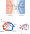
Figure 2. Ephrin B2/EphB4 signaling contributes to the fate of blood vessel identity
A. Schematic diagram showing an ephrinB2-expressing cell interacting with an EphB4-expressing cell. Interactions between the ligand and receptor cause activation and downstream signaling events in both cells. EphrinB2 signaling in endothelial cells contributes to an arterial fate for the cell, while EphB4 signaling results in a venous fate. B. Development of a mature vascular bed requires interaction between ephrinB2-expressing and EphB4-expressing cells. This interaction ensures the formation of a hierarchically-organized system consisting of both arteries and veins of various size as well as a well-defined capillary bed. Mice deficient in both ephrinB2 and EphB4 die in utero as a result of defective remodeling of the vascular bed. These mice contain primitive blood vessels that fail to develop into a mature vasculature. These mice have no distinguishable arteries or veins. Likewise, all vessels are of the same caliber and no capilliaries are present.
Figure 3
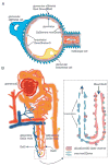
Figure 3. Heterogeneity within the renal vessels
A. Each glomerulus is surrounded by multiple layers of cells that serve as a specialized filtration barrier. Glomerular endothelial cells form the initial barrier to blood flowing through the kidney and are characterized by numerous fenestrations. B. The vessels of the vasa recta illustrate the complexity and precise specialization of the renal vascular system. The descending vasa recta are lined by a continuous endothelium that is punctuated by a high density of aquarporin-1 water channels and urea transporters (see insert). The ascending vasa recta are lined by a fenestrated endothelium (see insert) and have fewer conducting channels.
Figure 4
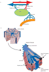
Figure 4. The liver has a dual blood supply
A. Schematic illustration of the arrangement of blood flow in the liver. Blood enters the liver via 2 major vessels: the hepatic artery (carrying oxygenated blood from the aorta) and the portal vein (carrying nutrient-rich blood from the intestine). Blood leaves the liver via the hepatic vein that subsequently drains into the inferior vena cava. B. The vascular arrangement within a liver lobule. Blood enters the lobule through a branch of the hepatic artery and portal vein and flows through the sinusoid to the central vein, which in turn empties into the hepatic vein that leaves the liver and drains into the vena cava.

References

    1. Risau W. Mechanisms of angiogenesis. Nature. 1997;386:671–674. - PubMed
    1. Klein R. Eph/ephrin signaling in morphogenesis, neural development and plasticity. Current Opinion in Cell Biology. 2004;16:580–589. - PubMed
    1. Kullander K, Klein R. Mechanisms and functions of eph and ephrin signalling. Nat Rev Mol Cell Biol. 2002;3:475–486. - PubMed
    1. Himanen JP, Chumley MJ, Lackmann M, Li C, Barton WA, Jeffrey PD, Vearing C, Geleick D, Feldheim DA, Boyd AW, Henkemeyer M, Nikolov DB. Repelling class discrimination: ephrin-A5 binds to and activates EphB2 receptor signaling. Nat Neurosci. 2004;7:501–509. - PubMed
    1. Wang H, Anderson DJ. Eph Family Transmembrane Ligands Can Mediate Repulsive Guidance of Trunk Neural Crest Migration and Motor Axon Outgrowth. Neuron. 1997;18:383–396. - PubMed

Publication types

Substances