

Loss of SR-A and CD36 activity reduces atherosclerotic lesion complexity without abrogating foam cell formation in hyperlipidemic mice
- PMID: 18948635
- PMCID: PMC2666043
- DOI: 10.1161/ATVBAHA.108.176644
Loss of SR-A and CD36 activity reduces atherosclerotic lesion complexity without abrogating foam cell formation in hyperlipidemic mice
Abstract
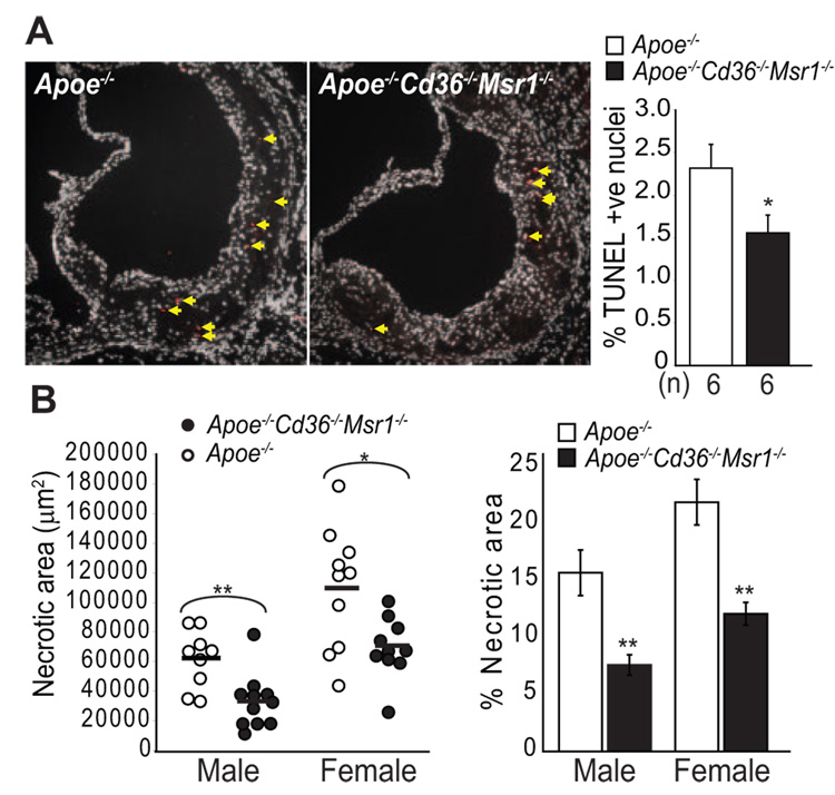
Objective: The scavenger receptors SR-A and CD36 have been implicated in macrophage foam cell formation during atherogenesis and in the regulation of inflammatory signaling pathways, including those leading to lesional macrophage apoptosis and plaque necrosis. To test the impact of deleting these receptors, we generated Apoe(-/-) mice lacking both SR-A and CD36 and fed them a Western diet for 12 weeks.
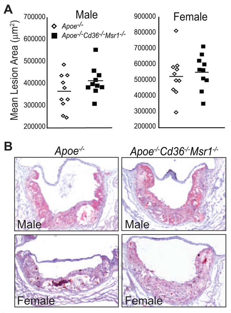
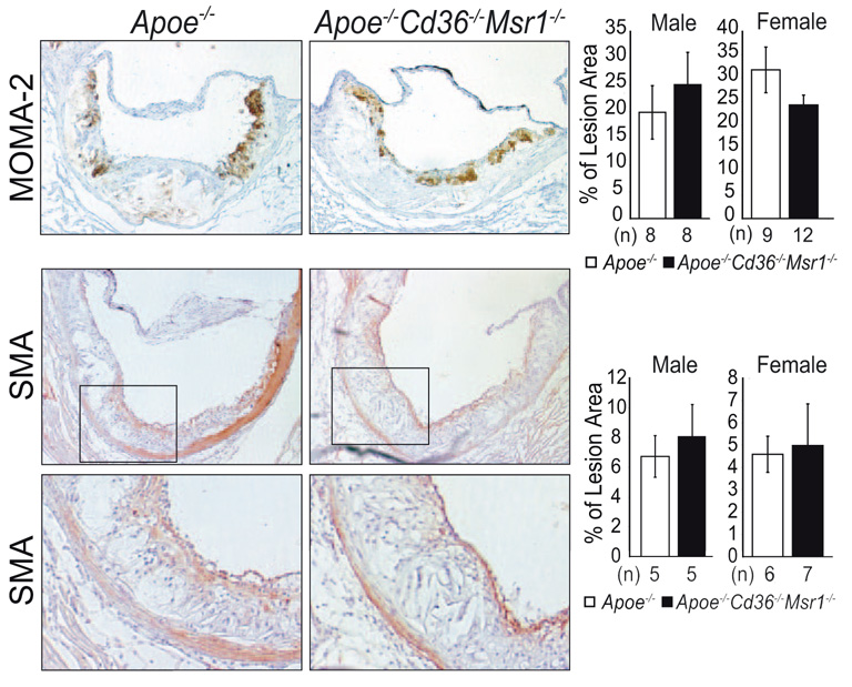
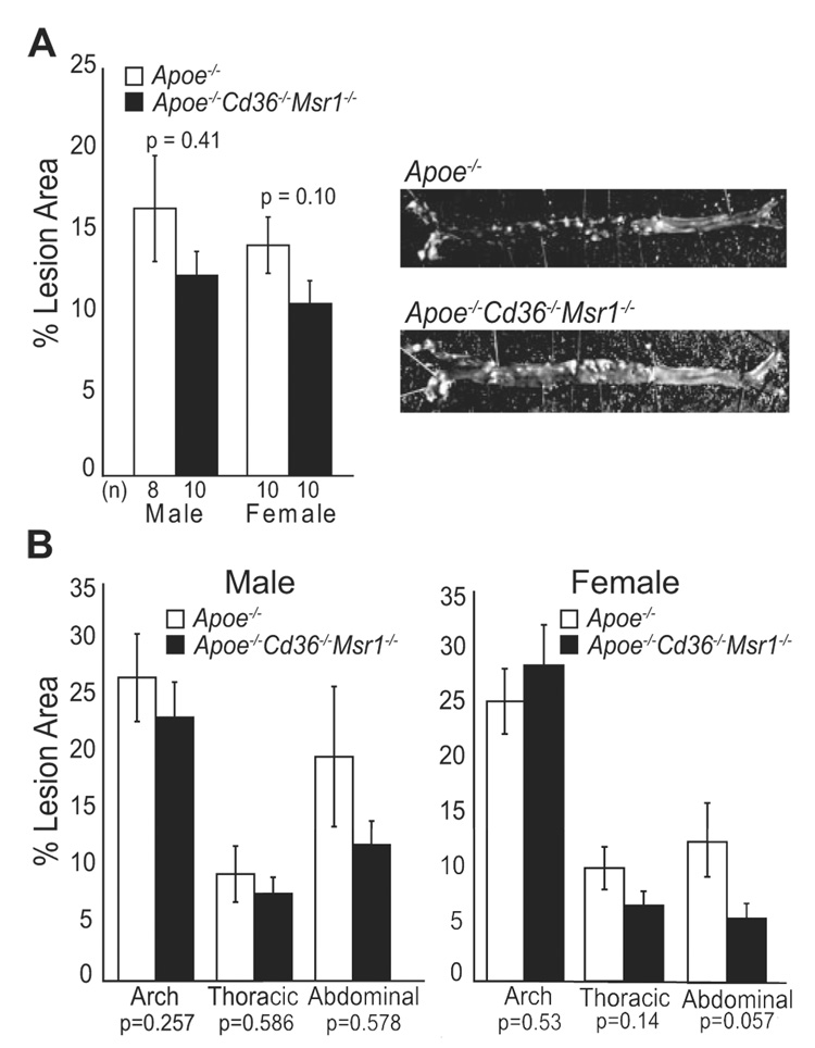
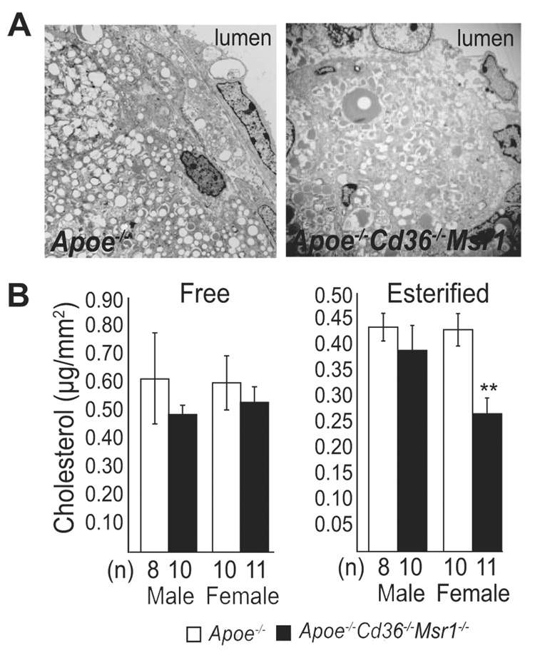
Methods and results: We analyzed atheroma in mice, assessing lesion size, foam cell formation, inflammatory gene expression, apoptosis, and necrotic core formation. Aortic root atherosclerosis in Apoe(-/-)Cd36(-/-)Msr1(-/-) mice, as assessed by morphometry, electron microscopy, and immunohistochemistry, showed no decrease in lesion area or in vivo foam cell formation when compared to Apoe(-/-) mice. However, Apoe(-/-)Cd36(-/-)Msr1(-/-) lesions showed reduced expression of inflammatory genes and morphological analysis revealed a approximately 30% decrease in macrophage apoptosis and a striking approximately 50% decrease in plaque necrosis in aortic root lesions of these mice.
Conclusions: Although targeted deletion of SR-A and CD36 does not abrogate macrophage foam cell formation or substantially reduce atherosclerotic lesion area in Apoe(-/-) mice, loss of these pathways does reduce progression to more advanced necrotic lesions. These data suggest that targeted inhibition of these pathways in vivo may reduce lesional inflammation and promote plaque stability.
Figures
References
-
- Moore KJ, Freeman MW. Scavenger receptors in atherosclerosis: beyond lipid uptake. Arterioscler Thromb Vasc Biol. 2006;26:1702–1711. - PubMed
-
- Tabas I, Williams KJ, Boren J. Subendothelial lipoprotein retention as the initiating process in atherosclerosis: update and therapeutic implications. Circulation. 2007;116:1832–1844. - PubMed
-
- Libby P, Geng YJ, Aikawa M, Schoenbeck U, Mach F, Clinton SK, Sukhova GK, Lee RT. Macrophages and atherosclerotic plaque stability. Curr Opin Lipidol. 1996;7:330–335. - PubMed
Publication types
MeSH terms
Substances
Grants and funding
LinkOut - more resources
Full Text Sources
Other Literature Sources
Medical
Molecular Biology Databases
Research Materials
Miscellaneous
