

Comment
doi: 10.1038/4551047a.
Apoptosis: Stabbed in the BAX
- PMID: 18948940
- PMCID: PMC3242476
- DOI: 10.1038/4551047a
Item in Clipboard
Comment
Apoptosis: Stabbed in the BAX
Nature.
.
Abstract
Apoptotic cell death is an intricate and highly regulated process. To initiate apoptosis, the protein BIM binds to a hitherto unrecognized site on the BAX protein to trigger permeabilization of the outer mitochondrial membrane.
Figures
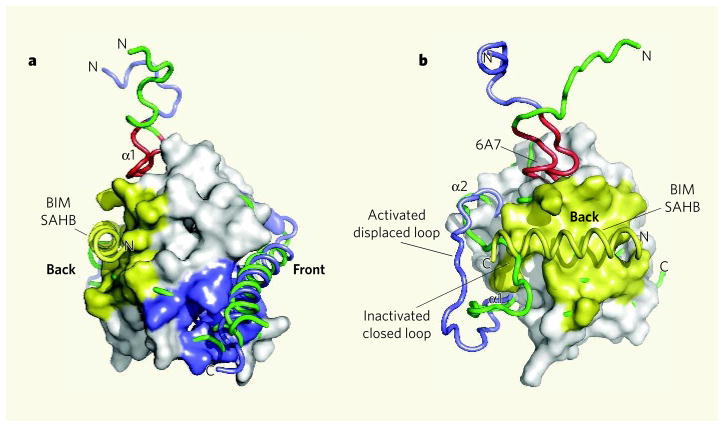
a, The structure of BAX based on a previous study is shown with BIM SAHB binding at a site on the ‘back’ of the molecule (yellow). This contrasts with the expected binding of a BH3-domain helix in the conventional (at least for anti-apoptotic proteins) hydrophobic BH pocket located at the ‘front’ of the molecule (blue). b, Walensky, Tjandra and colleagues find that conformational changes accompany the binding of a BIM SAHB peptide to BAX. The chain containing α-helix 1 (α1) and the loop between α1 and α2 is shown before (green) and after (blue) binding of BIM SAHB. The BAX amino-terminal region 6A7, which becomes exposed on association with BIM SAHB, is shown in red. (Images prepared by T. Moldoveanu, St Jude Children’s Research Hospital.)

a, On the basis of the latest structural information, we speculate that the binding of BIM SAHB to the ‘back’ of BAX, and the ensuing conformational changes, constrain the ‘front’ of the molecule, including the hydrophobic BH pocket, causing the carboxy-terminal region (not shown) and α-helix 4 containing the BH3 domain (green) to move out of the BH pocket and create a groove. Two such BAX molecules could then dimerize ‘nose-to-nose’ through interactions between the BH3 domain and the hydrophobic BH groove. b, Interaction at an additional, undefined interface might result in the formation of BAX oligomers and so permeabilization of the outer mitochondrial membrane. On BAX activation, the inducer of activation — in this case BIM SAHB, but presumably any activator — might dissociate to allow higher-order oligomer formation.
Comment on
-
BAX activation is initiated at a novel interaction site.Nature. 2008 Oct 23;455(7216):1076-81. doi: 10.1038/nature07396. Nature. 2008. PMID: 18948948 Free PMC article.
References
Publication types
MeSH terms
Substances
Grants and funding
LinkOut - more resources
Full Text Sources
Other Literature Sources
Research Materials
