. 2008 Oct;4(10):e1000236.
doi: 10.1371/journal.pgen.1000236. Epub 2008 Oct 24.

Two pathways recruit telomerase to Saccharomyces cerevisiae telomeres

Affiliations

Two pathways recruit telomerase to Saccharomyces cerevisiae telomeres

Angela Chan et al. PLoS Genet. 2008 Oct.

Abstract

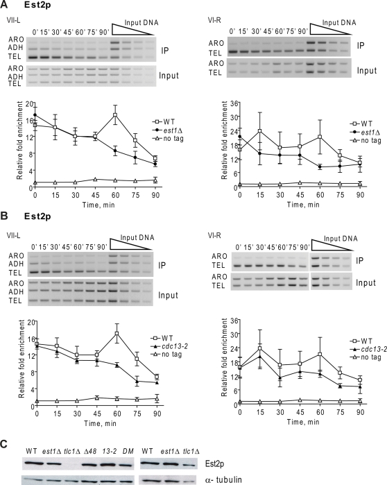
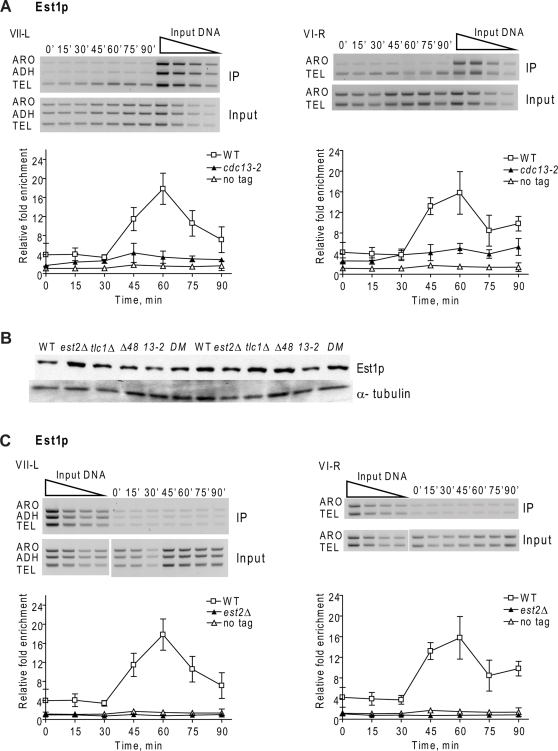
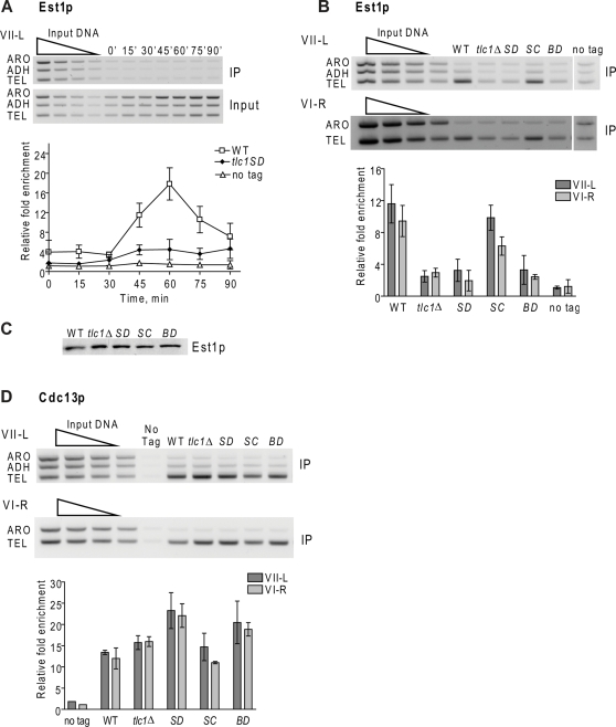
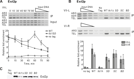
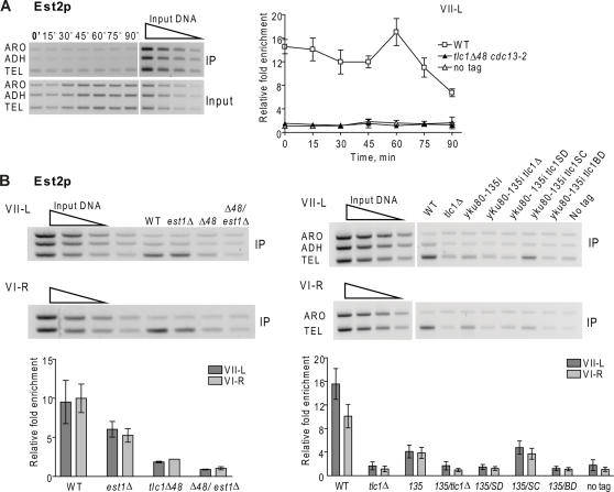
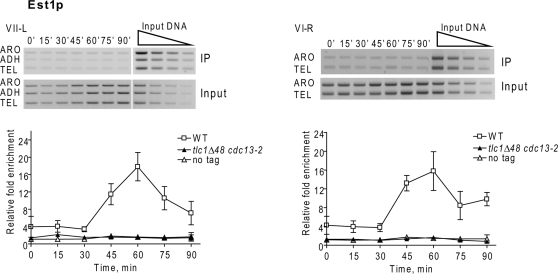
The catalytic subunit of yeast telomerase, Est2p, is a telomere associated throughout most of the cell cycle, while the Est1p subunit binds only in late S/G2 phase, the time of telomerase action. Est2p binding in G1/early S phase requires a specific interaction between telomerase RNA (TLC1) and Ku80p. Here, we show that in four telomerase-deficient strains (cdc13-2, est1A, tlc1-SD, and tlc1-BD), Est2p telomere binding was normal in G1/early S phase but reduced to about 40-50% of wild type levels in late S/G2 phase. Est1p telomere association was low in all four strains. Wild type levels of Est2p telomere binding in late S/G2 phase was Est1p-dependent and required that Est1p be both telomere-bound and associated with a stem-bulge region in TLC1 RNA. In three telomerase-deficient strains in which Est1p is not Est2p-associated (tlc1-SD, tlc1-BD, and est2A), Est1p was present at normal levels but its telomere binding was very low. When the G1/early S phase and the late S/G2 phase telomerase recruitment pathways were both disrupted, neither Est2p nor Est1p was telomere-associated. We conclude that reduced levels of Est2p and low Est1p telomere binding in late S/G2 phase correlated with an est phenotype, while a WT level of Est2p binding in G1 was not sufficient to maintain telomeres. In addition, even though Cdc13p and Est1p interact by two hybrid, biochemical and genetic criteria, this interaction did not occur unless Est1p was Est2p-associated, suggesting that Est1p comes to the telomere only as part of the holoenzyme. Finally, the G1 and late S/G2 phase pathways for telomerase recruitment are distinct and are likely the only ones that bring telomerase to telomeres in wild-type cells.


Conflict of interest statement

The authors have declared that no competing interests exist.

Figures

Figure 1
Figure 1. Est2p telomere binding in late S/G2 phase is reduced in est1Δ and cdc13-2 cells.
Cells expressing Est2-G8-Myc or lacking a Myc-tagged protein (no tag) were arrested in G1 phase. After release from the arrest, cells were grown at 24°C and samples taken at 15 min intervals for FACS and chromatin immuno-precipitation (ChIP). After DNA purification, PCR amplification was carried out with telomeric (TEL), subtelomeric (ADH), and non-telomeric (ARO) primers on DNA from immuno-precipitates (IP) or whole cell lysates (Input). Two-fold serial dilutions of input DNA established the linear range of the reactions (Input, top right). In this and subsequent figures, the agarose gels are representative data from mutant strains. For each time point, binding is expressed as the relative fold enrichment of TEL over ARO signal after normalization to input DNA. Error bars are ±1 standard deviations from ≥3 independent synchronies. A. Est2p binding to VII-L (left) and VI-R (right) telomeres in synchronous est1Δ (black circles) versus WT (white squares), or untagged (white triangles) cells. The values for Est2p binding to the VII-L telomere were not significantly different in WT versus est1Δ cells (P values >0.05), except at 60 (P value of 0.002), 75 (P = 0.02) and 90 (P = 0.04) minutes. Est2p binding to the VI-R telomere in WT versus est1Δ cells was significantly different only at 60 (P = 0.001) and 75 (P = 0.01) min. B. Est2p binding to VII-L (left) and VI-R (right) telomeres in synchronous cdc13-2 (black triangles) versus WT (white squares), or untagged (white triangles) cells. Est2p binding to the VII-L telomere were significantly different in WT versus cdc13-2 cells only at 60 (P = 0.01) and 75 (P = 0.02) minutes. At VI-R, binding was significantly different only at 75 minutes (P = 0.005) C. Western analyses of Est2p-G8-Myc or α-tubulin levels in extracts from WT and mutant strains with duplicate extracts prepared from independent colonies. The lanes labeled Δ48 are from tlc1Δ48 cells; 13-2 is cdc13-2; DM is double mutant tlc1Δ48 cdc13-2. Est2p-G8-Myc in lane 3 (tlc1Δ) was detectable upon longer exposure; see last lane that has protein sample from another tlc1Δ isolate and Figure 4C.
Figure 2
Figure 2. Est1p telomere binding is greatly reduced in synchronous cdc13-2 cells and eliminated in est2Δ cells.
Methods and symbols are as described in the Figure 1 legend except that cells expressed Est1-Myc. A. Est1p binding to VII-L (left) and VI-R (right) telomeres in synchronous cdc13-2 (black triangles) versus WT (white squares), or untagged (white triangles) cells. Est1p binding to the VII-L telomere was significantly higher in cdc13-2 cells than in the no-tag control at times of peak Est1p binding (P values ranged from 0.0003 at 60 min to 0.035 at 90 min). Est1p binding to the VI-R telomere was significantly different from the no-tag control at all time points (P values ranged from 0.0006 at 60 min to 0.0065 at 90 min.). B. Western analyses of Est1p-Myc versus α-tubulin levels in extracts from WT and mutant strains. The lanes labeled Δ48 are from different tlc1Δ48 colonies; 13-2 is cdc13-2; DM is double mutant tlc1Δ48 cdc13-2. C. Est1p binding to VII-L (left) and VI-R (right) telomeres in synchronous est2Δ (black triangles) versus WT (white squares), or untagged (white triangles) cells. Est1p binding to the VII-L and VI-R telomeres in est2Δ cells was modestly higher than the no-tag control only at 45 (P = 0.04 VII-L; 0.013, VI-R) and 60 min (P = 0.04, VII-L; 0.03, VI-R).
Figure 3
Figure 3. Est1p telomere binding is low in the absence of the stem-bulge region of TLC1 RNA.
Methods and symbols are as described in legend of Figure 1 except that cells expressed Est1-Myc and for panels B–D, asynchronous log phase cells were analyzed. A. Est1p binding to VII-L telomere in synchronous tlc1-SD (black diamonds) versus WT (white squares), or untagged (white triangles) cells. Although Est1p binding from 30 through 90 min was higher in tlc1-SD cells than in the no tag control, the difference was significant (P = 0.045) only at 45 min. B. Est1p binding to VII-L (top) or VI-R (bottom) telomeres in WT and mutant asynchronous cells. Bar graphs show average Est1p association with telomere VII-L (dark grey) or VI-R (light grey) with error bars indicating ±one standard deviation from that average; abbreviations for strains are SD, tlc1-SD; SC, tlc1-SC; BD, tlc1-BD. The level of binding in tlc1-SC cells was not significantly different from WT (P = 0.16, VII-L; 0.07, VI-R). The level of binding in both tlc1-SD and tlc1-BD cells was not significantly different from tlc1Δ cells (P values ranged from 0.22 to 0.29). The level of Est1p binding in tlc1Δ cells was significantly higher than in the no-tag control (P = 0.011, VII-L; 0.029, VI-R). Likewise, the level of Est1p binding in tlc1-SD (P = 0.03, VII-L; 0.13, VI-R) and tlc1-BD (P = 0.074, VII-L; 0.07, VI-R) was mostly significantly higher than the no-tag control. C. Western analyses of Est1p-Myc in extracts from WT and mutant strains. Abbreviations for strains are same as in panel B. D. Cdc13p binding to VII-L (top) and VI-R (bottom) telomeres in asynchronous mutant and WT cells. Bar graphs and symbols are as in panel B. Cdc13p binding was not significantly different in tlc1-SD versus tlc1-BD cells (P = 0.5, VII-L; 0.2, VI-R). Cdc13p binding was significantly higher than in WT at both telomeres in tlc1-SD (0.016,VII-L; 0.009,VI-R) and at VI-R in tlc1-BD (P = 0.015; but not at VII-L, P = 0.072). Cdc13p binding was similar in WT and tlc1Δ cells (P = 0.08, VII-L; 0.06, VI-R).
Figure 4
Figure 4. Est2p telomere binding in late S/G2 phase is reduced in mutants that lack the stem-bulge region of telomerase RNA.
Methods are the same as in Figure 1 legend. A. Est2p binding to VII-L telomere in synchronous tlc1-SD (black diamonds) versus WT (white squares), or untagged (white triangles) cells. Est2p binding to the VII-L telomere was significantly lower in tlc1-SD than in WT cells late in the cell cycle (from 45 to 90 min, P values ranged from P = 0.0007 at 60 min to 0.018 at 90 min). B. Est2p binding to VII-L (top) or VI-R (bottom) telomeres in asynchronous WT and mutant cells. Bar graphs show average Est2p association with telomere VII-L (dark grey) or VI-R (light grey) with error bars indicating ±one standard deviation from that average; abbreviations for strains are SD, tlc1-SD; SC, tlc1-SC; and BD, tlc1-BD. The level of Est2p telomere binding in tlc1-SD cells was significantly lower than in WT (P = 0.004, VII-L; 0.0003, VI-R) as well as at the VII-L telomere in tlc1-BD cells (P = 0.001). The level of Est2p telomere binding in WT and tlc1-SC cells was not significantly different (P = 0.083, VII-L; 0.573, VI-R). C. Western analysis of Est2p-G8-Myc in extracts from WT and mutant strains. Abbreviations for strains are same as in panel B.
Figure 5
Figure 5. Est2p telomere binding is eliminated in mutants lacking both the G1 and the late S/G2 phase recruitment pathways.
Methods are the same as in Figure 1 legend. A. Est2p binding to VII-L telomere in synchronous tlc1Δ48 cdc13-2 (black triangles) versus WT (white squares), or untagged (white triangles) cells. Est2p telomere binding in the double mutant was not different from the no-tag control (P = 0.11 to 0.85) except at the 0 min time point (P = 0.039). B. Est2p binding to VII-L (top) or VI-R (bottom) telomeres in asynchronous WT and mutant cells. Bar graphs show average Est2p association with telomere VII-L (dark grey) or VI-R (light grey). Abbreviations are Δ48, tlc1Δ48; 13-2, cdc13-2; 135, yku80-135i, SD, tlc1-SD; SC, tlc1-SC; BD, tlc1-BD. For double mutants, there is a slash between the two alleles as in Δ48/est1Δ which stands for tlc1Δ48 est1Δ. Error bars indicating ±one standard deviation from that average. Est2p binding was indistinguishable in tlc1Δ versus the double mutants yku80-135i tlc1-SD or yku80-135i tlc1-BD cells (P values ranged from 0.28 to 0.85) while Est2p binding in the double mutant yku80-135i tlc1-SC was significantly greater than in tlc1Δ (P = 0.003, VII-L; 0.0001, VI-R).
Figure 6
Figure 6. Est1p telomere binding is eliminated in mutants lacking both the G1 and the late S/G2 phase recruitment pathways.
Methods are the same as in Figure 1 legend. Est1p binding to VII-L (left) or VI-R (right) telomeres in synchronous tlc1Δ48 cdc13-2 (black triangles) versus WT (white squares), or untagged (white triangles) cells. Est1p telomere binding in the double mutant tlc1Δ48 cdc13-2 was indistinguishable from the no-tag control at all time points at the VII-L telomere (P ranged from 0.12 to 0.98) except at 15 min (P = 0.0007). Est1p binding in the double mutant was indistinguishable at the VI-R telomere (P values ranged from 0.38 to 0.64) except at 15 (P = 0.0423) and 30 (P = 0.0399) mins.

References

    1. Vega L, Mateyak M, Zakian V. Getting to the end: telomerase access in yeast and humans. Nat Rev Mol Cell Biol. 2003;4:948–959. - PubMed
    1. Lundblad V, Szostak JW. A mutant with a defect in telomere elongation leads to senescence in yeast. Cell. 1989;57:633–643. - PubMed
    1. Wellinger RJ, Wolf AJ, Zakian VA. Origin activation and formation of single-strand TG1–3 tails occur sequentially in late S phase on a yeast linear plasmid. Mol Cell Biol. 1993;13:4057–4065. - PMC - PubMed
    1. Wellinger RJ, Wolf AJ, Zakian VA. Saccharomyces telomeres acquire single-strand TG1–3 tails late in S phase. Cell. 1993;72:51–60. - PubMed
    1. Wellinger RJ, Ethier K, Labrecque P, Zakian VA. Evidence for a new step in telomere maintenance. Cell. 1996;85:423–433. - PubMed

Publication types