

Increased SPARC expression in primary angle closure glaucoma iris
- PMID: 18949063
- PMCID: PMC2571946
Increased SPARC expression in primary angle closure glaucoma iris
Abstract
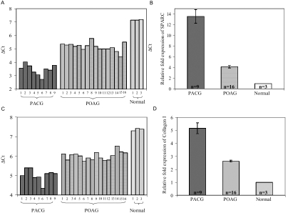
Purpose: SPARC (secreted protein, acidic, and rich in cysteine) is involved in extracellular matrix (ECM) organization. The purpose of this study was to evaluate the expression of SPARC in iris tissue from primary angle closure glaucoma (PACG) eyes.
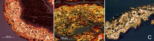
Methods: Iris tissue was obtained from peripheral iridectomies performed during trabeculectomy surgery in nine PACG and 16 primary open-angle glaucoma (POAG) eyes at the Singapore National Eye Centre. Three non-glaucoma control iris specimens were obtained from patients who underwent Descemet's stripping automated endothelial keratoplasty (DSAEK) procedure. SPARC and collagen I expression were quantified by real-time polymerase chain reaction (PCR). The histological distribution of collagen I and III in the iris stroma was determined using picrosirius red polarization. Density of the iris stromal vasculature was also calculated.
Results: The mean age was 68.9+/-10.9 years and 65.7+/-12.2 years in POAG and PACG groups, respectively. The PACG iris expressed SPARC 13.6-fold more and collagen I 5.2 fold more compared to non-glaucoma control iris. The PACG iris also demonstrated 3.3 fold higher SPARC and 2.0 fold higher collagen I expression relative to the POAG iris. The density of collagen I was greater in PACG eyes than in POAG and control eyes (p<0.001). The mean density of iris stromal blood vessels per micron square area was similar in all three groups.
Conclusions: SPARC was significantly increased in the PACG iris. The data suggest that SPARC could play a role in the development of PACG by influencing the biomechanical properties of the iris through a change in ECM organization.
Figures
Similar articles
-
Distinct iris gene expression profiles of primary angle closure glaucoma and primary open angle glaucoma and their interaction with ocular biometric parameters.Clin Exp Ophthalmol. 2016 Nov;44(8):684-692. doi: 10.1111/ceo.12743. Epub 2016 May 2. Clin Exp Ophthalmol. 2016. PMID: 26988898 Free PMC article.
-
Interocular asymmetry of visual field defects in primary open angle glaucoma and primary angle-closure glaucoma.Eye (Lond). 2004 Apr;18(4):365-8. doi: 10.1038/sj.eye.6700664. Eye (Lond). 2004. PMID: 15069431 Clinical Trial.
-
Expression of cyclooxygenase-1 and -2 in normal and glaucomatous human eyes.Invest Ophthalmol Vis Sci. 2001 Oct;42(11):2616-24. Invest Ophthalmol Vis Sci. 2001. PMID: 11581208
-
Genetic susceptibility to primary angle closure glaucoma (PACG).Discov Med. 2013 Jan;15(80):17-22. Discov Med. 2013. PMID: 23375010 Review.
-
Angle-closure glaucoma: the role of the lens in the pathogenesis, prevention, and treatment.Surv Ophthalmol. 2009 Mar-Apr;54(2):211-25. doi: 10.1016/j.survophthal.2008.12.002. Surv Ophthalmol. 2009. PMID: 19298900 Review.
Cited by
-
Correlation of iris collagen and in-vivo anterior segment structures in patients in different stages of chronic primary angle-closure in both eyes.Indian J Ophthalmol. 2019 Oct;67(10):1638-1644. doi: 10.4103/ijo.IJO_1406_18. Indian J Ophthalmol. 2019. PMID: 31546499 Free PMC article.
-
Distinct iris gene expression profiles of primary angle closure glaucoma and primary open angle glaucoma and their interaction with ocular biometric parameters.Clin Exp Ophthalmol. 2016 Nov;44(8):684-692. doi: 10.1111/ceo.12743. Epub 2016 May 2. Clin Exp Ophthalmol. 2016. PMID: 26988898 Free PMC article.
-
The genetic mechanisms of primary angle closure glaucoma.Eye (Lond). 2015 Oct;29(10):1251-9. doi: 10.1038/eye.2015.124. Epub 2015 Jul 24. Eye (Lond). 2015. PMID: 26206529 Free PMC article. Review.
-
Updates on Genes and Genetic Mechanisms Implicated in Primary Angle-Closure Glaucoma.Appl Clin Genet. 2021 Mar 9;14:89-112. doi: 10.2147/TACG.S274884. eCollection 2021. Appl Clin Genet. 2021. PMID: 33727852 Free PMC article. Review.
-
Matrix metalloproteinases and tissue inhibitors of metalloproteinases in the aqueous humour of patients with primary angle closure glaucoma - a quantitative study.BMC Ophthalmol. 2014 Mar 24;14:33. doi: 10.1186/1471-2415-14-33. BMC Ophthalmol. 2014. PMID: 24655889 Free PMC article.
References
-
- Huang EC, Barocas VH. Active iris mechanics and pupillary block: steady-state analysis and comparison with anatomical risk factors. Ann Biomed Eng. 2004;32:1276–85. - PubMed
-
- Ningli W, Wenbin Z, Tiancai Y, Qiang Y, Mingying L, Meihua L. Studies of primary angle closure glaucoma in China. Yan Ke Xue Bao. 1997;13:120–4. - PubMed
-
- Konstas AG, Marchall GE, Lee WR. Immunocytochemical localisation of collagens (1-V) in the human iris. Graefes Arch Clin Exp Ophthalmol. 1990;228:180–6. - PubMed
Publication types
MeSH terms
Substances
LinkOut - more resources
Full Text Sources
Other Literature Sources
Miscellaneous