. 2007:1:1-7.
doi: 10.2174/1874192400701010001. Epub 2007 Jun 11.

Carotid Body AT(4) Receptor Expression and its Upregulation in Chronic Hypoxia

Affiliations

Carotid Body AT(4) Receptor Expression and its Upregulation in Chronic Hypoxia

Man-Lung Fung et al. Open Cardiovasc Med J. 2007.

Abstract

Hypoxia regulates the local expression of angiotensin-generating system in the rat carotid body and the me-tabolite angiotensin IV (Ang IV) may be involved in the modulation of carotid body function. We tested the hypothesis that Ang IV-binding angiotensin AT(4) receptors play a role in the adaptive change of the carotid body in hypoxia. The expression and localization of Ang IV-binding sites and AT(4) receptors in the rat carotid bodies were studied with histochemistry. Specific fluorescein-labeled Ang IV binding sites and positive staining of AT(4) immunoreactivity were mainly found in lobules in the carotid body. Double-labeling study showed the AT(4) receptor was localized in glomus cells containing tyrosine hydroxylase, suggesting the expression in the chemosensitive cells. Intriguingly, the Ang IV-binding and AT(4) immunoreactivity were more intense in the carotid body of chronically hypoxic (CH) rats (breathing 10% oxygen for 4 weeks) than the normoxic (Nx) control. Also, the protein level of AT(4) receptor was doubled in the CH comparing with the Nx group, supporting an upregulation of the expression in hypoxia. To examine if Ang IV induces intracellular Ca(2+) response in the carotid body, cytosolic calcium ([Ca(2+)](i)) was measured by spectrofluorimetry in fura-2-loaded glomus cells dissociated from CH and Nx carotid bodies. Exogenous Ang IV elevated [Ca(2+)](i) in the glomus cells and the Ang IV response was significantly greater in the CH than the Nx group. Hence, hypoxia induces an upregulation of the expression of AT(4) receptors in the glomus cells of the carotid body with an increase in the Ang IV-induced [Ca(2+)]i elevation. This may be an additional pathway enhancing the Ang II action for the activation of chemoreflex in the hypoxic response during chronic hypoxia.

Keywords: Angiotensin IV; angiotensin IV receptor; chemoreceptor; type-I cells.


Figures

Fig. (1)
Fig. (1)
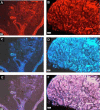
Angiotensin IV (Ang IV)-binding sites in normoxic (Nx) and chronically hypoxic (CH) rat carotid body. Panels on top show FITC-coupled Ang IV binding (A), + Ang IV (B), + Sarile (C) in Nx group. Panels in middle show FITC-coupled Ang IV binding (D), + Ang IV (E), + Sarile (F) in CH group. Panels at bottom show FITC-coupled Ang IV binding (G), + Ang IV (H), + Sarile (I) in rat kidney, which was used as the positive control. Concentration of FITC-coupled Ang IV, unlabeled Ang IV and Sarile were 0.2, 10, 1 μM, respectively. Calibration bar is 20 μm.
Fig. (2)
Fig. (2)
The expression and localization of AT4 receptor-immunoreactivity in (A) Nx and (B) CH rat carotid body. Note the intensity of staining is at a high level in the CH comparing with that of the Nx group. Sections with tyrosine hydroxylase (TH) for the Nx and CH carotid bodies are shown in C and D. E and F are overlays of the AT4 receptor- and TH-stainings. Calibration bars are 20 and 50 μm, respectively, for low (A, C, E) and high magnification (B, D, F).
Fig. (3)
Fig. (3)
Protein expression of AT4 receptors in Nx and CH rat carotid body. Note the level of protein expression increased significantly in the CH group. The expression in the hippocampus serves as the positive control. Densitometric analysis depicts the relative expression of AT4 receptor protein in the CH group of carotid bodies and the expression is normalized as percent of the Nx group. Statistical significance (n=5, p<0.05, unpaired-t test) is shown in asterisk when comparing with the Nx group.
Fig. (4)
Fig. (4)
The [Ca2+]i response to exogenous Ang IV in dissociated glomus cells of the rat carotid body from Nx control (A) and CH group (B). The records were made in cluster of 8-10 cells loaded with fura-2. Ang IV-induced [Ca2+]i elevation was more significant in the CH glomus cells than that of the Nx group. Note that the transient [Ca2+]i increase following the injection of Ang IV (0.01-10 μM) into the bath. In addition, histotoxic hypoxia with sodium cyanide (NaCN, 1 mM) increased the [Ca2+]i of the cells. Results are summarized in (C). On average, the [Ca2+]i elevation (ΔR (340/380)) induced by 10 μM Ang IV in the CH group (n=9) was significantly greater than that of the Nx control (n=7). Statistical significance (p<0.05, unpaired-t test) is shown in asterisk when comparing with the other groups.

Similar articles

Cited by

References

    1. Gonzalez C, Almaraz L, Obeso A, Rigual R. Carotid body chemoreceptors: from natural stimuli to sensory discharges. Physiol Rev. 1994;74:829–98. - PubMed
    1. Lahiri S, Rozanov C, Roy A, Storey B, Buerk DG. Regulation of oxygen sensing in peripheral arterial chemoreceptors. Int J Biochem Cell Biol. 2001;33:755–74. - PubMed
    1. Biscoe TJ, Duchen MR, Eisner DA, O’Neill SC, Valdeolmillos M. Measurements of intracellular Ca2+ in dissociated type I cells of the rabbit carotid body. J Physiol. 1989;416:421–34. - PMC - PubMed
    1. Buckler KJ, Vaughan-Jones RD. Effects of hypercapnia on membrane potential and intracellular calcium in rat carotid body type I cells. J Physiol. 1994;478(Pt 1):157–71. - PMC - PubMed
    1. Dasso LL, Buckler KJ, Vaughan-Jones RD. Interactions between hypoxia and hypercapnic acidosis on calcium signaling in carotid body type I cells. Am J Physiol Lung Cell Mol Physiol. 2000;279:L36–42. - PubMed

LinkOut - more resources