. 2008 Oct;49(10):1963-75.
doi: 10.1080/10428190802322919.

Inhibition of p38alpha MAPK disrupts the pathological loop of proinflammatory factor production in the myelodysplastic syndrome bone marrow microenvironment

Affiliations

Inhibition of p38alpha MAPK disrupts the pathological loop of proinflammatory factor production in the myelodysplastic syndrome bone marrow microenvironment

Tony Navas et al. Leuk Lymphoma. 2008 Oct.

Erratum in

  • Leuk Lymphoma. 2009 Sep;50(9):1554. Bhagat, T [added]; Gajavelli, S [added]; Kambhampati, S [added]

Abstract

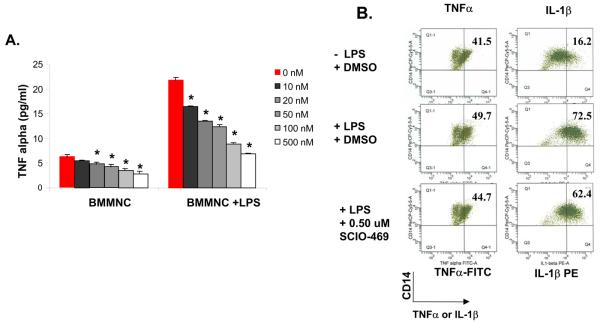
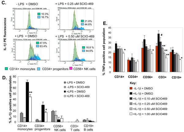
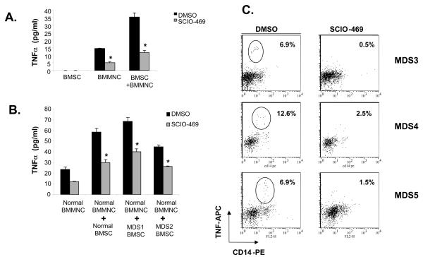
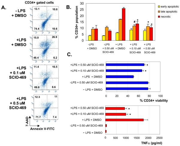
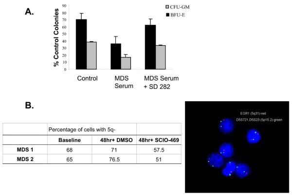
Myelodysplastic syndromes (MDS) are common causes of ineffective hematopoiesis and cytopenias in the elderly. Various myelosuppressive and proinflammatory cytokines have been implicated in the high rates of apoptosis and hematopoietic suppression seen in MDS. We have previously shown that p38 MAPK is overactivated in MDS hematopoietic progenitors, which led to current clinical studies of the selective p38alpha inhibitor, SCIO-469, in this disease. We now demonstrate that the myelosuppressive cytokines TNFalpha and IL-1beta are secreted by bone marrow (BM) cells in a p38 MAPK-dependent manner. Their secretion is stimulated by paracrine interactions between BM stromal and mononuclear cells and cytokine induction correlates with CD34+ stem cell apoptosis in an inflammation-simulated in vitro bone marrow microenvironment. Treatment with SCIO-469 inhibits TNF secretion in primary MDS bone marrow cells and protects cytogenetically normal progenitors from apoptosis ex vivo. Furthermore, p38 inhibition diminishes the expression of TNFalpha or IL-1beta-induced proinflammatory chemokines in BM stromal cells. These data indicate that p38 inhibition has anti-inflammatory effects on the bone marrow microenvironment that complements its cytoprotective effect on progenitor survival. These findings support clinical investigation of p38alpha as a potential therapeutic target in MDS and other related diseases characterised by inflammatory bone marrow failure.


Figures

Figure 1
Figure 1. Inflammatory bone marrow mononuclear cells secrete TNFα and IL-1β in a p38 MAPK-dependent manner
A. Bone marrow mononuclear cells (BMMNC) (1 × 10⁶) from a normal healthy donor were cultured in the absence or presence of increasing concentrations of SCIO-469 for 24h without or with 10 ng/mL LPS. TNFα concentration in cell supernatants was determined by ELISA. Figure represents Mean ± SD of three independent experiments. *P < 0.01 vs “DMSO”. B. Primary BM-derived CD14+ cells from a normal donor were incubated in IMDM + 10% FBS in the presence or absence of 20 ng/mL LPS and SCIO-469 for 4h. Brefeldin A (golgi plug) was added to a final concentration of 2 ug/mL during the last hour of incubation. Cells were harvested, washed with FBS staining buffer and labeled with anti-CD14-PerCP Cy5.5 followed by intracellular staining with anti-IL-1β–PE and anti-TNFα–FITC. Figure shows percent double-stained CD14+ TNFα+ (left) and CD14+ IL-1β+ (right) in the same cell population. C. BMMNC from a normal donor (1 × 10⁶) were incubated in the presence or absence of 10 ng/mL LPS for 4h. Brefeldin A (golgi plug) was added to a final concentration of 2 ug/mL during the last hour of incubation. Cells were harvested, washed and labeled with different fluorochrome-conjugated antibodies to CD14 (monocytes), CD56 (NK cells) and CD34 (progenitor cells) followed by intracellular staining with PE-conjugated anti-IL-1β. Figure shows the relative IL-1β expression for each of the specific BM populations: CD14+ cells (green), CD34+ cells (light blue), CD56+ cells (violet). D BMMNC from a different normal donor (1 × 10⁶) were treated with or without 0.5 μM SCIO-469 and incubated in the presence or absence of 10 ng/mL LPS for 4h. Brefeldin A (golgi plug) was added to a final concentration of 2 ug/mL during the last hour of incubation. Cells were harvested, washed and labeled with different fluorochrome-conjugated antibodies to CD45 (leukocytes), CD14, CD3 (T cells), CD19 (B cells), CD56 and CD34 followed by intracellular staining with PE-conjugated anti-IL-1β. Figure shows the relative IL-1β expression for each of the specific BM population. Results are expressed as Mean ± SD of three independent experiments. **P < 0.001 or *P < 0.01 vs “+ LPS − SCIO-469”. E. BMMNC (1 × 10⁶) were incubated without or with increasing concentrations of SCIO-469 and in the presence or absence of 50 ng/mL IL-1β for 24h. Brefeldin A was added to a final concentration of 2 ug/ml during the last 2 hours of incubation. Cells were harvested, washed, labeled and then fixed with different fluorochrome-conjugated antibodies to CD45, (leukocytes), CD14 (monocytes), CD3 (T cells), CD19 (B cells), CD56 (NK cells) and CD34 (progenitor cells) followed by intracellular staining with PE-conjugated anti-TNFα. Figure shows the relative TNFα expression for each of the specific BM populations. Results are expressed as mean +/− S.D. of three independent experiments. **P < 0.001 or *P < 0.01 or #P < 0.05 vs “+ IL-1β − SCIO-469”.
Figure 2
Figure 2. Secretion of TNF requires p38 MAPK-dependent interactions between stromal and mononuclear cells
A. BMSC and BMMNC from normal donors were either cultured alone or cocultured together for 72h in the presence and absence of 0.5 μM SCIO-469. TNFα concentration in cell supernatants was determined by ELISA. Figure represents Mean ± SD of three independent experiments. B. Similar co-culture experiments were conducted as in (A) but using BMSC derived from either normal healthy control or from low risk MDS patients. Figure represents Mean ± SD of three independent experiments. C. Total bone marrow aspirates isolated from three different MDS patients were assessed for intracellular TNFα production by using the Cytofix/Cytoperm kit according to the manufacturer's instructions. Briefly, cells were incubated in the presence of 10 μg/mL of immobilized anti-CD3 mAb plus 2 μg/mL of anti-CD28 antibody with or without 0.5 μM SCIO-469 in the presence of monensin for 6h at 37°C. Addition of anti-CD28 antibody providing costimulation and monensin blocking secretion of the cytokines during the incubation allows sensitive assessment of cytokine-producing cells. After the stimulation, cells were stained with anti-CD14-PE and TNFα-APC before analyzing by flow cytometry. CD14+ cells with intracellular TNF were expressed as a percentage of total CD14+ cells.
Figure 3
Figure 3. SCIO-469 inhibits LPS-induced CD34+ Apoptosis and TNFα production in normal BMMNC in vitro
A. BMMNC (1 × 10⁶) from a normal healthy donor were cultured in the absence or presence of increasing concentrations of SCIO-469 without or with 10 ng/mL LPS for 48h. Cells were stained with anti-CD34-PE Cy7, anti-CD45-APC Cy7, Annexin V-PE and 7-AAD and analyzed by flow cytometry using the BD LSR II. Dot plot shows Annexin V-PE (X-axis) and 7-AAD (Y-axis) staining of CD34+ gated cells. B. Bar graph showing percent early apoptotic (Annexin V+, 7-AAD−), late apoptotic/necrotic (Annexin V+, 7-AAD+), and necrotic (Annexin V−, 7-AAD+) in CD34+ gated cell populations. Figures represents Mean ± SD of three independent experiments. *P < 0.01 or #P < 0.05 vs “+ LPS − SCIO-469”. C. TNF concentration was measured by ELISA in supernatants collected from experiment performed above and correlated with percentage of viable CD34+ cells. Figures represent Mean ± SD of three independent experiments. *P < 0.01 for DMSO + LPS.
Figure 4
Figure 4. SCIO-469 can protect normal stem cell clones from apoptosis in MDS
A. Normal CD34+ cells obtained from healthy volunteer were grown in methylcelluose in the presence 100 μL of primary bone marrow sera obtained from bone marrows of 3 MDS patients. These experiments were done in the presence or absence of 100 nM SD-282. Erythroid and Myeloid colonies were counted after 14 days. Mean ± SD of 3 independent experiments are shown. B. BMMNC from 2 patients with MDS with chromosome 5q deletion were cultured in the presence or absence of 0.5 μM SCIO-469 for 48h. Cells were fixed onto slides pre- and post-treatment and used for fluorescent in situ hybridization using EGR-1 probe (5q31-RED) to detect the number of abnormal clones. A 5p15 centromeric (GREEN) probe was used as internal control. 200 cells per slide were counted and result expressed as percentage.

Similar articles

Cited by

References

    1. Greenberg P, Cox C, LeBeau MM, Fenaux P, Morel P, Sanz G, Sanz M, Vallespi T, Hamblin T, Oscier D. International scoring system for evaluating prognosis in myelodysplastic syndromes. Blood. 1997;89:2079–88. and others. - PubMed
    1. Heaney ML, Golde DW. Myelodysplasia. N Engl J Med. 1999;340:1649–60. - PubMed
    1. Verma A, List AF. Cytokine targets in the treatment of myelodysplastic syndromes. Curr Hematol Rep. 2005;4:429–35. - PubMed
    1. Allampallam K, Shetty V, Mundle S, Dutt D, Kravitz H, Reddy PL, Alvi S, Galili N, Saberwal GS, Anthwal S. Biological significance of proliferation, apoptosis, cytokines, and monocyte/macrophage cells in bone marrow biopsies of 145 patients with myelodysplastic syndrome. Int J Hematol. 2002;75:289–97. and others. - PubMed
    1. Claessens YE, Park S, Dubart-Kupperschmitt A, Mariot V, Garrido C, Chretien S, Dreyfus F, Lacombe C, Mayeux P, Fontenay M. Rescue of early stage myelodysplastic syndrome-deriving erythroid precursors by the ectopic expression of a dominant negative form of FADD. Blood. 2005 - PubMed

Publication types

MeSH terms