A preliminary fMRI study of analgesic treatment in chronic back pain and knee osteoarthritis
- PMID: 18950528
- PMCID: PMC2584040
- DOI: 10.1186/1744-8069-4-47
A preliminary fMRI study of analgesic treatment in chronic back pain and knee osteoarthritis
Abstract
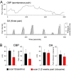
The effects of an analgesic treatment (lidocaine patches) on brain activity in chronic low back pain (CBP) and in knee osteoarthritis (OA) were investigated using serial fMRI (contrasting fMRI between before and after two weeks of treatment). Prior to treatment brain activity was distinct between the two groups: CBP spontaneous pain was associated mainly with activity in medial prefrontal cortex, while OA painful mechanical knee stimulation was associated with bilateral activity in the thalamus, secondary somatosensory, insular, and cingulate cortices, and unilateral activity in the putamen and amygdala. After 5% lidocaine patches were applied to the painful body part for two weeks, CBP patients exhibited a significant decrease in clinical pain measures, while in OA clinical questionnaire based outcomes showed no treatment effect but stimulus evoked pain showed a borderline decrease. The lidocaine treatment resulted in significantly decreased brain activity in both patient groups with distinct brain regions responding in each group, and sub-regions within these areas were correlated with pain ratings specifically for each group (medial prefrontal cortex in CBP and thalamus in OA). We conclude that the two chronic pain conditions involve distinct brain regions, with OA pain engaging many brain regions commonly observed in acute pain. Moreover, lidocaine patch treatment modulates distinct brain circuitry in each condition, yet in OA we observe divergent results with fMRI and with questionnaire based instruments.
Figures


References
-
- Deyo RA, Weinstein JN. Low back pain. N Engl J Med. 2001;344:363–370. - PubMed
-
- Apkarian AV, Sosa Y, Krauss BR, Thomas PS, Fredrickson BE, Levy RE, et al. Chronic pain patients are impaired on an emotional decision-making task. Pain. 2004;108:129–136. - PubMed
-
- Grachev ID, Fredrickson BE, Apkarian AV. Abnormal brain chemistry in chronic back pain: an in vivo proton magnetic resonance spectroscopy study. Pain. 2000;89:7–18. - PubMed
Publication types
MeSH terms
Substances
Grants and funding
LinkOut - more resources
Full Text Sources
Medical

