. 2010 Sep;31(9):1503-15.
doi: 10.1016/j.neurobiolaging.2008.08.019. Epub 2008 Oct 23.

Beta amyloid and hyperphosphorylated tau deposits in the pancreas in type 2 diabetes

Affiliations

Beta amyloid and hyperphosphorylated tau deposits in the pancreas in type 2 diabetes

Judith Miklossy et al. Neurobiol Aging. 2010 Sep.

Abstract

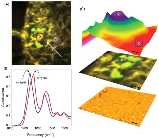
Strong epidemiologic evidence suggests an association between Alzheimer disease (AD) and type 2 diabetes. To determine if amyloid beta (Abeta) and hyperphosphorylated tau occurs in type 2 diabetes, pancreas tissues from 21 autopsy cases (10 type 2 diabetes and 11 controls) were analyzed. APP and tau mRNAs were identified in human pancreas and in cultured insulinoma beta cells (INS-1) by RT-PCR. Prominent APP and tau bands were detected by Western blotting in pancreatic extracts. Aggregated Abeta, hyperphosphorylated tau, ubiquitin, apolipoprotein E, apolipoprotein(a), IB1/JIP-1 and JNK1 were detected in Langerhans islets in type 2 diabetic patients. Abeta was co-localized with amylin in islet amyloid deposits. In situ beta sheet formation of islet amyloid deposits was shown by infrared microspectroscopy (SIRMS). LPS increased APP in non-neuronal cells as well. We conclude that Abeta deposits and hyperphosphorylated tau are also associated with type 2 diabetes, highlighting common pathogenetic features in neurodegenerative disorders, including AD and type 2 diabetes and suggesting that Abeta deposits and hyperphosphorylated tau may also occur in other organs than the brain.


Conflict of interest statement

Disclosure

The authors have no actual or potential interest to disclose.

Figures

Fig. 1
Fig. 1
Expression of APP and tau. mRNAs in human pancreas tissue, SH-SY5Y and beta cell lines. (A) Ethidium bromide-stained agarose gel of RT-PCR products from human pancreas of 5 individuals, 3 with type 2 diabetes (lanes 1–3) and 2 controls (lanes 4 and 5). Molecular weight markers are shown on the right and left side. (B) Expression of APP and tau mRNAs in pancreas tissue of three additional diabetic (lanes 1, 3, 5) and one control (lane 7) patients compared to the frontal cortex of the same patients (lanes 2, 4, 6, 8, respectively). (C) The expression of APP and tau mRNAs in cultured INS-1 beta cells was compared to those of neuroblastoma SH-SY5Y cells. Ethidium bromide-stained agarose gel of RT-PCR products from beta cells (lane 1) compared to SH-SY5Y cells (lane 2). Molecular weight markers are shown on the left side. Glyceraldehyde 3-phosphate dehydrogenase (GAPDH) was used to verify loading conditions.
Fig. 2
Fig. 2
Detection of APP and tau in human pancreas tissue by Western blot. Immunoblotting using a monoclonal anti-APP antibody (clone C2212) which recognizes a common N terminal epitope of the three major APP isoforms detects immunoreactive APP bands around 120 kDa in three pancreas samples (lanes 1–2 correspond to controls and lane 3 to type 2 diabetes) when compared to N2a human neuroblastoma cells which are known to contain APP and tau and which were used as positive controls (lane 4). Two tau reactive bands of ca. 64 and 69 kDa were detected with anti-tau, clone tau 2 antibody in the same pancreas samples similar to those of N2a neuroblastoma cells.
Fig. 3
Fig. 3
Islet amyloid deposit in type 2 diabetes. (A) Section of the pancreas from a patient with type 2 diabetes stained with H&E. Arrows point to a Langerhans islet, the site of amyloid deposition in type 2 diabetes, which is surrounded by acinar cells. At high magnification (B) a cell with “flame”-like triangular shape (arrow) and homogeneous eosinophilic cytoplasm is visible. (C) Islet amyloid deposits exhibiting positive fluorescence, localized in Langerhans islets in a case with type 2 diabetes as stained with the fluorochrome Thioflavin S which detects amyloid. (D) Amylin immunoreactive amyloid deposits localized to degenerating Langerhans islets in the pancreas of a patient with type 2 diabetes. Rabbit anti-amylin antibody raised against islet amyloid peptide (rabbit anti-IAPP 1–37) was used for immunostaining. Bar: (A) 150 μm; (B) 7 μm; (C) 150 μm; (D) 200 μm.
Fig. 4
Fig. 4
Aβ in islet amyloid deposits in type 2 diabetes. (A) Islet amyloid deposits showing positive Aβ immunoreaction with anti-Aβ antibodies 21F12 (A), 2F9AF (B) and 4G8 (C) which recognize different epitopes of the molecule (see Table 1). (D and E) Sections of the pancreas from a diabetic patient immunostained with rabbit anti-amylin antibody (D, IAPP 1–37, Dr. A. Clark) and with the 4G8 monoclonal anti-Aβ antibody (E) showing their intra-cellular localization (arrows). (F) Small group of affected acinar cells showing Aβ immunoreaction demonstrated with the anti-Aβ monoclonal antibody 21F12. (G–I) Pancreas section of a patient with type 2 diabetes doubly immunostained with an anti-amylin monoclonal antibody (GTX 74673, GeneTex, Inc.) labeled with TRITC-tagged secondary anti-mouse antibody (which gives a red fluorescence for amylin) (G) and with a polyclonal antibody to the C terminus of Aβ 40) (Dr. H. Mori) which was labeled with a FITC-tagged anti-rabbit secondary antibody shows a green fluorescence. The orange color of the merged image shows the co-localization of amylin and Aβ in islet amyloid deposits. Bars: (A and C) 250 μm, (B) 70 μm, (D and E) 50 μm, (F) 200 μm, (G–I) 120 μm.
Fig. 5
Fig. 5
Hyperphosphorylated tau in the pancreas in type 2 diabetes. (A–C) Pancreas sections of patient with type 2 diabetes showing a positive immunoreaction to tau A0024 (A and B) and AT8 (C). Positive immunoreaction in the affected Langerhans islet to Ubiquitin (D), apo-E (E), Apo(a) (F), IB1/JIP-1 (G) and JNK1 (H). (I) Control section where the primary antibody was replaced by normal serum. Bars: (A and C) 250 μm, (B) 70 μm, (D) 200 μm, (E–I) 100 μm.
Fig. 6
Fig. 6
Synchrotron infrared microspectroscopy (SIRMS) analysis of islet amyloid deposits. (A) Epifluorescence image showing Thioflavin S positive amyloid deposits (arrow). (B and C) Infrared absorption image of protein structure (1630/1655 cm−1) showed an absorbance maximum near 1630 cm−1 representative of β-sheet protein structure. In areas of the pancreas without Thioflavin S stained amyloid, the absorbance maximum was near 1655 cm−1 corresponding to α-helical protein structure. Scale bar: 10 μm.
Fig. 7
Fig. 7
LPS increases APP levels in PC12 and TE671 cells. Western blot analysis of PC12 and TE671 cells using anti-APP 22C11 antibody detected increased APP levels following 6 and 24 and 48 h of LPS exposure. The increased APP levels were comparable in PC12 and TE671 cells.
Fig. 8
Fig. 8
Western blot analysis of APP levels in PC12 and TE671 cells following exposure to insulin. Decreased APP levels were detected following 6–48 h of exposures to human recombinant insulin 100 ng/ml exposures in PC12 and in non-neuronal TE671 cells.

References

    1. Alafuzoff I, Aho L, Helisalmi S, Mannermaa A, Soininen H. β-Amyloid deposition in brains of subjects with diabetes. Neuropathol Appl Neurobiol. 2008 doi: 10.1111/j.1365-2990.2008.00948.x. - DOI - PubMed
    1. Arvanitakis Z, Wilson RS, Bienias JL, Evans DA, Bennett DA. Diabetes mellitus and risk of Alzheimer disease and decline in cognitive function. Arch Neurol. 2004;61:661–666. - PubMed
    1. Arvanitakis Z, Schneider JA, Wilson RS, Li Y, Arnold SE, Wang Z, Bennett DA. Diabetes is related to cerebral infarction but not to AD pathology in older persons. Neurology. 2006;67:1960–1965. - PubMed
    1. Asfari M, Janjic D, Meda P, Li G, Halban PA, Wollheim CB. Establishment of 2-mercaptoethanol-dependent differentiated insulin-secreting cells. Endocrinology. 1992;130:167–178. - PubMed
    1. Askanas V, Engel WK, Bilak M, Alvarez RB, Selkoe DJ. Twisted tubulofilaments of inclusion body myositis muscle resemble paired helical filaments of Alzheimer brain and contain hyperphosphorylated tau. Am J Pathol. 1994;144:177–187. - PMC - PubMed

Publication types