. 2008 Oct 24;32(2):259-75.
doi: 10.1016/j.molcel.2008.10.002.

Atomic structure of the KEOPS complex: an ancient protein kinase-containing molecular machine

Affiliations

Atomic structure of the KEOPS complex: an ancient protein kinase-containing molecular machine

Daniel Y L Mao et al. Mol Cell. .

Abstract

Kae1 is a universally conserved ATPase and part of the essential gene set in bacteria. In archaea and eukaryotes, Kae1 is embedded within the protein kinase-containing KEOPS complex. Mutation of KEOPS subunits in yeast leads to striking telomere and transcription defects, but the exact biochemical function of KEOPS is not known. As a first step to elucidating its function, we solved the atomic structure of archaea-derived KEOPS complexes involving Kae1, Bud32, Pcc1, and Cgi121 subunits. Our studies suggest that Kae1 is regulated at two levels by the primordial protein kinase Bud32, which is itself regulated by Cgi121. Moreover, Pcc1 appears to function as a dimerization module, perhaps suggesting that KEOPS may be a processive molecular machine. Lastly, as Bud32 lacks the conventional substrate-recognition infrastructure of eukaryotic protein kinases including an activation segment, Bud32 may provide a glimpse of the evolutionary history of the protein kinase family.


Figures
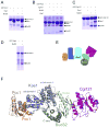
Figure 1
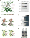
Figure 1. Architecture of the KEOPS complex
(A–D) GST pull down experiments with purified KEOPS subunits. Proteins were separated by SDS-PAGE and visualized by Coomassie blue staining. (E) Architectural model of the KEOPS complex derived from (A–D). (F) Composite model of the KEOPS complex based on the X-ray crystal structures of single subunits, binary complexes and a ternary complex summarized in Table 1.
Figure 2
Figure 2. Structure and catalytic function of Bud32
(A) Ribbons representation of Bud32 showing important catalytic residues. For all figures and tables, residues of the archaeal ortholog are listed first and when appropriate, that of the S. cerevisiae ortholog are bracketed. Residue labels are colored according to the effect of the indicated mutations on growth in yeast (see color legend). The N-lobe of Bud32 is colored light green and the C-lobe is colored dark green. A metal ion and ATP were modeled into the catalytic cleft based on the structure of RIO1 (PDB 1ZP9). (B) Autophosphorylation analysis of Bud32 wild type or mutant proteins in the presence or absence of Cgi121. Assays were carried out with [γ-32P]-ATP at 60°C and stopped at the indicated time points. Products of the reactions were separated by SDS-PAGE and 32P incorporation was detected by autoradiography. (C) Ribbons comparison of Bud32 with Rio1 (PDB 1ZP9; top panel) and PKA (PDB 2CPK; bottom panel). Bud32 is colored as in (A) and Rio kinase is shown in red with its unique N-terminal lobe insertion (labeled as ‘Rio insert’) in orange. PKA is colored yellow with its A-loop shown in red. (D) Effect of Bud32 catalytic cleft mutations on the growth of yeast. A bud32Δ yeast strain was transformed with a centromeric low copy vector expressing wild type BUD32, the indicated Bud32 mutant or the empty vector as control. Liquid cultures of each strain were spotted on minimal medium in a series of 10 fold dilutions. Growth of yeast at 30°C was monitored over a 6 day period. Representative data is shown. A summary of doubling times in liquid culture for a selection of mutants is provided in Table S3. (E) Bud32 catalytic mutants have short telomeres. Terminal restriction fragment (TRF) analysis of the yeast strains described in (D).
Figure 3
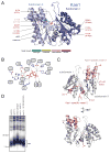
Figure 3. Structure and catalytic function of Kae1
(A) Ribbons representation of Kae1 highlighting catalytic cleft residues. Subdomains I and II are colored dark and light blue respectively. The metal ion and ADP were modeled into the cleft based on the structure of P. abyssi Kae1 (PDB 2IVP). (B) Schematic of catalytic cleft residues of Kae1 that coordinate ATP and metal ion (based on PDB 2IVP). (C) Kae1 has two characteristic inserts (colored pink and red) that distinguish it from other ASKHA fold proteins. Secondary structure elements within the unique inserts are labeled. (D) The kae1-E176R (E213R) mutant has short telomeres. TRF analysis was performed on the kae1Δ strain transformed with a low copy vector encoding wild type Kae1 or the E176 (E213R) mutant. Telomeres from wild type, bud32Δ and cgi121Δ strains were used as reference.
Figure 4
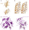
Figure 4. Structures of Pcc1 and Cgi121
(A) Ribbons representation of the Pcc1 monomer (top) and dimer (bottom). Pcc1 protomers 1 and 2 are colored light and dark orange respectively. (B) Stereo view of conserved hydrophobic contacts within the Pcc1 dimer interface. (C) Ribbons representation of human Cgi121 (left) and M.jannaschii Cgi121 (right) (rmsd=1.92 Å).
Figure 5
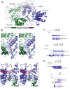
Figure 5. Binding mode of Bud32 to Kae1
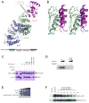
(A) Ribbons representation of the Bud32-Kae1 complex. Kae1 and Bud32 are colored as in Figures 2A and 3A. Interface residues whose mutation gives rise to growth phenotypes in yeast are shown in stick representation. (B) Stereo zoom-in view of the Kae1-Bud32 binding interface centered on (top) Kae1-specific insert 1 and (bottom) C-terminal tail region of Bud32. Figures are colored as in (A). (C,D) Mutation of the Kae1-Bud32 binding interface. Binding experiments with the indicated GST fusion baits and prey proteins were performed as in Figure 1. (C) Examination of the Bud32-Kae1 interaction. Lane 1 (lower panel) is a loading control for Bud32. (D) Examination of the Cgi121-Bud32 interaction using Bud32 mutants (top) and the Pcc1-Kae1 interaction using Kae1 mutants (bottom).
Figure 6
Figure 6. Binding mode of Cgi121 to Bud32
(A) Ribbons representation of the Cgi121-Bud32-Kae1 complex. Bud32, Kae1 and Cgi121 are colored as in Figures 2A, 3A and 4B, respectively. Residues whose mutation gives rise to strong phenotypes in yeast are highlighted. (B) Stereo zoom-in view of the Bud32-Cgi121 binding interface showing side chains that participate in the interaction. (C) In vitro binding experiments with the indicated GST fusion baits and purified prey proteins. (D) Disruption of Cgi121 binding to Bud32 impairs the ability of Cgi121 to potentiate Bud32 autophosphorylation activity in vitro. Bud32 kinase reactions were performed as in Figure 2B. (E) Functional analysis in yeast of Bud32-Cgi121 binding interface mutations on cell growth. cgi121Δ yeast was transformed with empty vector, a vector encoding wild type Cgi121 or the L134E/A138R (L172E/I176R) double mutant. All strains were plated by serial dilution on minimal medium as described in Figure 2D. (F) Analysis of Cgi121 and Bud32 stability in vivo. Yeast encoding galactose-inducible 3xHA-Cgi121 and Bud32-FLAG proteins were grown for 4 hrs in media containing 2% glucose or 2% galactose. Cells grown in galactose were washed and resuspended in 2% glucose-containing media to repress the GAL1 promoter. Cells were harvested at times following glucose addition. α-HA and α-FLAG immunoblots are shown.
Figure 7
Figure 7. Binding mode of Pcc1 to Kae1
(A) Ribbons representation of the binding interface between Pcc1 and Kae1. Pcc1 and Kae1 are colored as in Figures 3 and 4, respectively. Residues whose mutation gives rise to a phenotype in yeast are highlighted. See Figure S13A for a zoom-in view of the Pcc1-Kae1 binding interface with participating side chains. (B–D) Mutational analysis of the Pcc1-Kae1 binding interface. Binding experiments with the indicated GST fusion baits and prey proteins were performed as in Figure 1. (B) Examination of various Kae1 proteins binding to Pcc1 (upper panel) or Bud32 (lower panel). (C) Examination of various Pcc1 proteins binding to Kae1. (D) Input gel shows equivalent quantities of soluble wild type and mutant Pcc1 proteins used in the binding reactions in (C). (E) Analysis of Kae1-Pcc1 interface mutations on yeast growth. Left panel: Serial dilutions of thermosensitive pcc1-4 strains harboring plasmid encoding wild type Pcc1, pcc1 mutants or empty vector were grown at 37°C (non-permissive) or 23°C (permissive) for 3–5 days. Right panel: kae1Δ yeast harboring plasmid encoding wild type Kae1, Kae1 mutants or empty vector were grown for 3–6 days.

References

    1. Abdullah KM, Lo RY, Mellors A. Cloning, nucleotide sequence, and expression of the Pasteurella haemolytica A1 glycoprotease gene. J Bacteriol. 1991;173:5597–5603. - PMC - PubMed
    1. Arigoni F, Talabot F, Peitsch M, Edgerton MD, Meldrum E, Allet E, Fish R, Jamotte T, Curchod ML, Loferer H. A genome-based approach for the identification of essential bacterial genes. Nat Biotechnol. 1998;16:851–856. - PubMed
    1. Bax A, Grzesiek S. Methodological Advances in Protein Nmr. Accounts of Chemical Research. 1993;26:131–138.
    1. Cornilescu G, Delaglio F, Bax A. Protein backbone angle restraints from searching a database for chemical shift and sequence homology. J Biomol NMR. 1999;13:289–302. - PubMed
    1. Dar AC, Dever TE, Sicheri F. Higher-order substrate recognition of eIF2alpha by the RNA-dependent protein kinase PKR. Cell. 2005;122:887–900. - PubMed

Publication types

MeSH terms