. 2008 Dec 26;384(4):780-97.
doi: 10.1016/j.jmb.2008.10.013. Epub 2008 Oct 14.

Three-dimensional reconstruction of tarantula myosin filaments suggests how phosphorylation may regulate myosin activity

Affiliations

Three-dimensional reconstruction of tarantula myosin filaments suggests how phosphorylation may regulate myosin activity

Lorenzo Alamo et al. J Mol Biol. .

Abstract

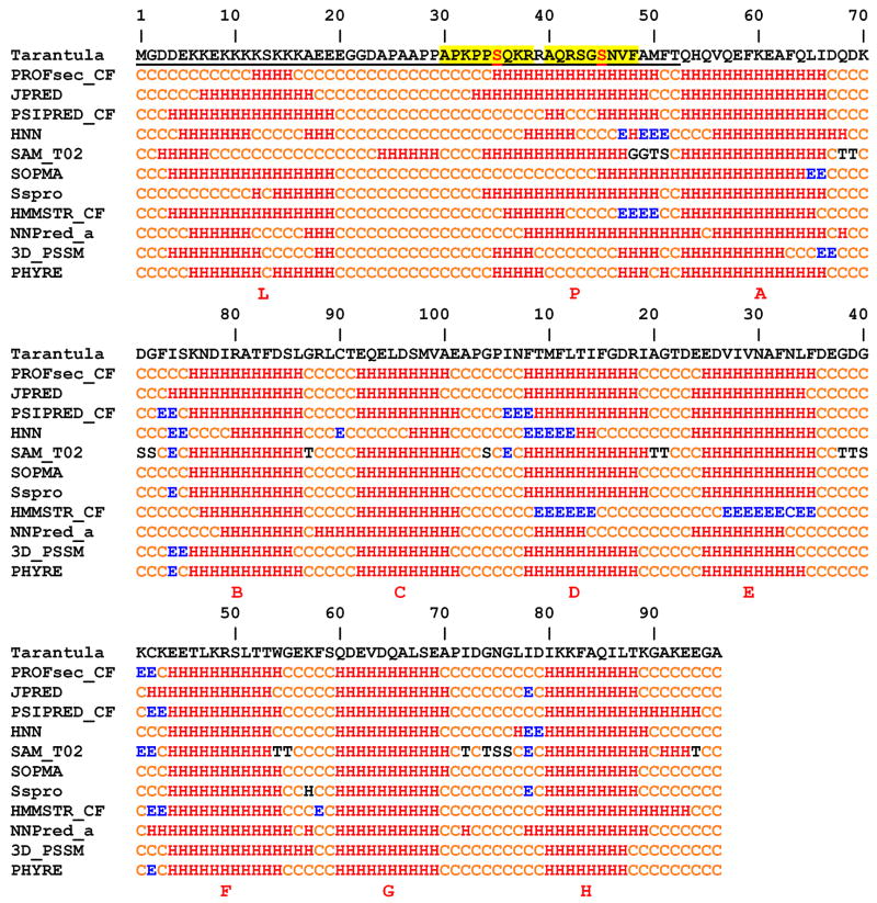
Muscle contraction involves the interaction of the myosin heads of the thick filaments with actin subunits of the thin filaments. Relaxation occurs when this interaction is blocked by molecular switches on these filaments. In many muscles, myosin-linked regulation involves phosphorylation of the myosin regulatory light chains (RLCs). Electron microscopy of vertebrate smooth muscle myosin molecules (regulated by phosphorylation) has provided insight into the relaxed structure, revealing that myosin is switched off by intramolecular interactions between its two heads, the free head and the blocked head. Three-dimensional reconstruction of frozen-hydrated specimens revealed that this asymmetric head interaction is also present in native thick filaments of tarantula striated muscle. Our goal in this study was to elucidate the structural features of the tarantula filament involved in phosphorylation-based regulation. A new reconstruction revealed intra- and intermolecular myosin interactions in addition to those seen previously. To help interpret the interactions, we sequenced the tarantula RLC and fitted an atomic model of the myosin head that included the predicted RLC atomic structure and an S2 (subfragment 2) crystal structure to the reconstruction. The fitting suggests one intramolecular interaction, between the cardiomyopathy loop of the free head and its own S2, and two intermolecular interactions, between the cardiac loop of the free head and the essential light chain of the blocked head and between the Leu305-Gln327 interaction loop of the free head and the N-terminal fragment of the RLC of the blocked head. These interactions, added to those previously described, would help switch off the thick filament. Molecular dynamics simulations suggest how phosphorylation could increase the helical content of the RLC N-terminus, weakening these interactions, thus releasing both heads and activating the thick filament.


Figures

Fig. 1
Fig. 1
3D-reconstruction of the frozen-hydrated tarantula thick filament, filtered to 2 nm resolution (see suppl. fig. 3 and suppl. methods), showing four helices of interacting-head motifs (one in yellow). The 3D-map segment shows four 14.5 crowns of interacting-heads. Bar 14.5 nm.
Fig. 2
Fig. 2
First line: Amino acid sequence of tarantula RLC. The 52 aa NTF is underlined. Lines 2–12: Prediction of secondary structure using the programs PROFsec, JPRED, PSIPRED, HNN, SAM, SOPMA, Sspro, HMMSTR, NNPred, 3D_PSSM and PHYRE. All programs predict ten helices, A–H corresponding to four EF hands motifs and L and P located in the NTF. Helix L, rich in positively charged Lys residues, is connected through a Pro-Ala repeat coil linker to the phosphorylation helix P, containing the two phosphorylatable serines (in red, Ser-35, Ser-45) each located in a MLCK consensus sequence (highlighted in yellow). H = helix, E = strand, C = coil, G = 3/10 helix, T = H-bonded turn and S = bend.
Fig. 3
Fig. 3
(a) Sequence alignment of RLCs for the 10 reported species with long NTFs. The two tarantula phosphorylatable serines (Ser-35 and Ser-45) are marked with arrows, and their MLCK consensus sequences with red boxes. UniProtKB ID code sequences (striated): Bombyx mori (Q1HPS0), Gryllotalpa orientalis (Q49M29), Riftia pachyptila (Q9GUA2), Clonorchis sinensis (Q2YHG1), Schistosoma japonicum (Q5DB55), Aedes aegypti (Q17HX1), Culex pipiens (Q45FA2), Anophelex gambae (Q7PUV3) and Drosophila melanogaster (P18432). (b) Sequence alignment of S2 from human cardiac and chicken smooth muscles. The S2 kink positions are indicated with red arrows: Met-877 (human36) and His-888 (chicken32). The black boxes show the two negatively charged ring 1 (Glu-905 – Asp-917) and ring 2 (Glu-932 – Gln-946) of S2.
Fig. 4
Fig. 4
Predicted atomic structure for the tarantula RLC obtained using the PredictProtein server. The structure has three domains: domain 1 (helices A–D), domain 2 (helices E–H) and domain 3 (helices L and P). The predicted secondary structure for the 52 aa NTF (domain 3 or “phosphorylation domain”13) is formed by a positively charged helix L and helix P (with phosphorylatable Ser-35 and Ser-45), connected by a Pro-Ala coil linker. The 8.5 nm IQ motif helix of the myosin heavy chain (Glu-812 – Phe-855) is shown in light gray.
Fig. 5
Fig. 5
(a) Starting HMM atomic model used for flexible fitting built using the predicted structure of the tarantula RLC (fig. 4), the structure of human S2 and the chicken smooth muscle HMM atomic model (without the S2). The kink (red spheres) in the S2 was located at Met-888, in agreement with Blankenfeldt et al. 2006 (see fig. 3b and 5c). The connected skeleton of 31 positional markers used for flexible fitting, located inside the density subset of the interacting-heads, is shown with gray spheres and rods. (b) Final HMM atomic model after the flexible fitting. The RLC of the blocked and free heads are shown in yellow and red, with their corresponding NTFs in tan (blocked) and pink (free). The ELC of the blocked and the free heads are shown in orange and purple. The heavy chain of the blocked and free head are shown in green and blue. The 3D-map is shown as a pale gray surface in (a) and (b). (c) A comparison of the crystal structure of the human S2 before (red α-helices) and after (blue and green α-helices) the flexible fitting, which only changed the position of the coil-coiled α-helices. The arrow head indicates that the position of the kink (Met-888) does not changed before and after the flexible fitting. C-terminus are Lys-974 and Leu-972 (top). N-terminus is Pro-849 (red spheres, bottom). The connection of the two α-helices of the S2 with the two α-helices of the interacting-heads regulatory domains was done between Ser-853 and Gln-852. The connection of the S2 N-terminal with the C-terminal structure of the heads required some unbending and torsion in the 6 (blocked head) or 8 (free head) intervening aa’s (see Results). (d) The nine interactions of the interacting-head motif in a relaxed thick filament, as viewed (towards) the filament surface (cf. 5a and b from above the filament surface). The five intramolecular interactions (a, d, e, f, g) are between: (a) the CM loop of the free head motor domain (blue) and the S2 of the interacting-head motif (blue); (d) the blocked head motor domain (green) and the free head motor domain (blue), (e) the blocked head motor domain (green) and the free head ELC (purple), (f) the blocked head motor domain (green) and the S2 of the interacting-head motif (blue and green), and (g) the blocked head ELC (orange) and the S2 of the interacting-head motif (blue). Four intermolecular interactions (b/b′, c/c′, h) are between : (b/b′) the blocked head RLC NTF (b, tan; b′, gold) and the I loop of the motor domain of the neighbor free head (b, gold, b′, blue); (c/c′) the blocked head ELC (c, orange, c′, gold) and the C loop of the neighboring free head (c, gold, c′, blue); (h) the SH3 of the blocked head motor domain (green) with the S2 of the neighbor interacting-head (pale blue backbone surface). An interaction between the blocked head ELC (orange) and the backbone is not shown. The 3D-map is shown as a pale blue (backbone S2) or gold surface (up and low interacting-head pairs).
Fig. 6
Fig. 6
(a) Stereo-pair of flexible atomic fitting of two adjacent interacting-heads on one helical track of the tarantula filament. Three interactions are labeled, an intramolecular interaction ‘a’, and intermolecular interactions ‘b’ and ‘c’. The 3D-map is shown as a pale gray surface. (b) Stereo-pair of intramolecular interaction ‘a’ between the CM or cardiomyopathy loop (yellow ribbon) of free head (Ile-407 – Val-425 highlighted using balls and sticks, Arg-411 residue is in yellow) with its own S2 (blue a-helix), intermolecular interaction ‘b’ between the NTF (tan) of the RLC (yellow) of the blocked head with the neighbour motor domain free head I loop (Leu-305 – Gln-327 shown in balls and sticks, green ribbon), and intermolecular interaction ‘c’ between the ELC of the blocked head (orange) and the C loop (Gln-361 – Asp-380 shown in balls and sticks, red ribbon) neighbour motor domain free head (blue). Glu-932 – Glu-946 of the negatively charged ring 2 of S2 (fig. 3b) are shown as balls and sticks. Ser-35 and Ser-45 are shown as pale blue and pale magenta spheres, respectively. (c) Stereo-pair of a comparison of the blocked (green) and free (yellow) heads (fig. 5b) from tarantula myosin, with the published crystal structures known as the pre-power stroke closed (1BR1) (red) and transition structures (1DFL) (pale yellow), and the near(1DFK)/post(2MYS)/like(2OY6) rigor (pink/light blue/purple) or detached internally uncoupled (1B7T) (dark blue) open structures. The structures of the blocked and free heads used for the flexible fitting were assumed initially to be in the closed conformation (fig. 5a). For clarity the ELC and RLC have been removed. Note that the regulatory domain a-helices for the 1BR1, 1DFL, and the blocked and the free head are in the same plane. The difference between the blocked and the free head regulatory domain a-helices is mostly a change in angular orientation.
Fig. 7
Fig. 7
The structure of the tarantula RLC in the atomic model following flexible fitting. The top row a–c shows the flexed RLCs (in ribbons) from the blocked (a, yellow/orange), blocked and free (b) and free (c, red/pink) heads. The NTF structures are asymmetric: the blocked head NTF (a, orange) exhibits a more compact conformation than the free head (c, pink). The second, third and fourth rows show the RLC electrostatic surface charges (blue positive, red negative) for the whole RLCs (d–f), the RLC without their NTFs (g–i) and the NTFs alone (j–l). The bottom row (m,n) shows the elongated NTF (m, ribbon; n, surface charge; arrows: Ser-35, Ser-45). In relaxed muscle the NTF of the dephosphorylated blocked head RLC (a,d) exhibits a compact conformation (j) that enables the interactions shown in fig. 5d, while the NTF of the basally Ser-35 phosphorylated free head RLC (c,f) is partially elongated (l). Both positively charged helices L are packed –due to complementary charges (see suppl. movie 1)- against the blocked head domain 1 (cf, see discussion). If phosphorylation in Ser-45 of the free head RLC produces elongation of its NTF (m,n), this could release the free head, exposing the blocked head NTF, which in turn could be phosphorylated at Ser-45, elongated (m,n) and the blocked head released. This mechanism could explain how RLC phosphorylation weakens the head-head interactions, releasing them so they can interact with the thin filament.
Figure 8
Figure 8
Five of the reported mutations in the myosin head heavy chain and S2 associated with FHC. Myosin heavy chain: R403Q, R403L or R403W (orange sphere) in the CM loop (yellow); and E924K (grey spheres), E927K (pink spheres), E930K (cyan spheres) and E935K (yellow spheres) in the negatively charged ring 2 of S2, facing the CM loop. The side chain orientations at the interface of the loop may be unreliable as this is not a structure determination down to atomic detail, but a prediction at amino acid level of detail. See legend of fig. 6b.

References

    1. Craig R, Padrón R. Molecular Structure of the Sarcomere. In: Engel AG, Franzini-Armstrong C, editors. Myology. McGraw-Hill, Inc; New York: 2004. pp. 129–166.
    1. Lehman W, Szent-Gyorgyi AG. Regulation of muscular contraction. Distribution of actin control and myosin control in the animal kingdom. J Gen Physiol. 1975;66:1–30. - PMC - PubMed
    1. Vibert P, Craig R, Lehman W. Steric-model for activation of muscle thin filaments. J Mol Biol. 1997;266:8–14. - PubMed
    1. Poole KJ, Lorenz M, Evans G, Rosenbaum G, Pirani A, Craig R, Tobacman LS, Lehman W, Holmes KC. A comparison of muscle thin filament models obtained from electron microscopy reconstructions and low-angle X-ray fibre diagrams from non-overlap muscle. J Struct Biol. 2006;155:273–284. - PubMed
    1. Szent-Gyorgyi AG. Regulation by myosin: how calcium regulates some myosins, past and present. Adv Exp Med Biol. 2007;592:253–264. - PubMed

Publication types

Associated data

LinkOut - more resources