. 2009 Feb;136(2):694-704.e4.
doi: 10.1053/j.gastro.2008.09.015. Epub 2008 Sep 18.

T cell-derived lymphotoxin regulates liver regeneration

Affiliations

T cell-derived lymphotoxin regulates liver regeneration

Alexei V Tumanov et al. Gastroenterology. 2009 Feb.

Abstract

Background & aims: The ability of the liver to regenerate hepatic mass is essential to withstanding liver injury. The process of liver regeneration is tightly regulated by distinct signaling cascades involving components of the innate immune system, cytokines, and growth factors. However, the role of the adaptive immune system in regulation of liver regeneration is not well-defined. The role of adaptive immune system in liver regeneration was investigated in lymphocyte-deficient mice and in conditional lymphotoxin-deficient mice.

Methods: A model of liver regeneration after 70% partial hepatectomy was used, followed by examination of liver pathology, survival, DNA synthesis, and cytokine expression.

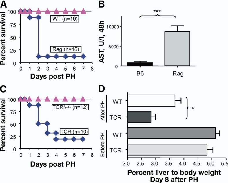
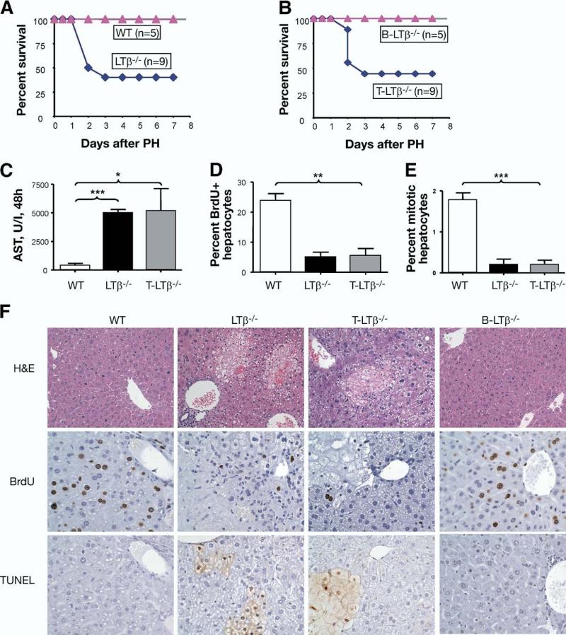
Results: We found that mice deficient in T cells show a reduced capacity for liver regeneration following partial hepatectomy. Furthermore, surface lymphotoxin, provided by T cells, is critical for liver regeneration. Mice specifically deficient in T-cell lymphotoxin had increased liver damage and a reduced capacity to initiate DNA synthesis after partial hepatectomy. Transfer of splenocytes from wild-type but not lymphotoxin-deficient mice improved liver regeneration in T cell-deficient mice. We found that an agonistic antibody against the lymphotoxin beta receptor was able to facilitate liver regeneration by reducing liver injury, increasing interleukin-6 production, hepatocyte DNA synthesis, and survival of lymphocyte-deficient (Rag) mice after partial hepatectomy.

Conclusions: The adaptive immune system directly regulates liver regeneration via a T cell-derived lymphotoxin axis, and pharmacological stimulation of lymphotoxin beta receptor might represent a novel therapeutic approach to improve liver regeneration.


Figures

Figure 1
Figure 1
T cells are essential for liver regeneration. Mice underwent 70% partial hepatectomy (PH), and survival was monitored for 8 days. Data represent 1 of 2 independent experiments. N = number of mice per group. (A) Rag mice show an increased mortality after PH compared with WT. (B) Increased liver damage in Rag mice after PH. Serum aspartate aminotransferase (AST) levels at 48 hours after PH. (C) T cell-deficient mice (TCRβδ–/–, designated TCR) show an increased mortality after PH. Mice with inactivation of TCRδ chain only (TCRδ–/–) show normal survival. (D) T cell-deficient mice show reduced ratio of liver to body mass at 8 days after PH.
Figure 2
Figure 2
Both CD8 and CD4 T cells are required for liver regeneration. (A and B) Mice depleted of CD8 or CD4 T cells show increased mortality after PH. Wild-type mice were injected with anti-CD8 or anti-CD4 antibody 24 hours prior to PH. (C and D) CD8–/– and CD4–/– mice show an increased mortality after PH. (E) CD8–/– mice display increased areas of liver necrosis and apoptosis and reduced DNA synthesis at 48 hours after PH. Liver sections were stained with H&E (left panel), anti-BrdU antibody (middle panel), and apoptotic cells using TUNEL protcol (right panel). Represents 1 of 2 independent experiments (5 mice per group). Original magnification, ×200. (F) Reduced DNA synthesis in CD8–/– mice at 48 hours after PH as determined by positive BrdU incorporation.
Figure 3
Figure 3
Adoptive splenocyte transfer improves liver regeneration in T cell-deficient mice. Fifty million splenocytes (Spl) from WT or LTα–/– mice were transferred to TCR mice 1 week prior to PH. Represents 1 of 2 independent experiments (5 mice per group). (A) ALT levels in serum at 48 hours after partial hepatectomy. (B) Increased DNA synthesis in T cell-deficient mice after transfer of splenocytes from WT but not from LTα-deficient mice. (C) Liver H&E sections at 48 hours after PH indicating the decreased injury in TCR mice reconstituted with WT but not LTα–/– splenocytes. Original magnification, ×400.
Figure 4
Figure 4
LT expressed by T cells is essential for liver regeneration. (A) Reduced survival of LTβ–/– mice after PH. (B) Increased mortality of T cell-specific LTβ-deficient mice (T-LTβ–/–) after PH. N = number of mice per group. (C) Increased liver damage of T-LTβ–/– mice after PH. AST levels in serum of indicated mice at 48 hours after partial hepatectomy are shown. Represents 1 of 2 independent experiments, 5 mice per group. (A–C) Represent 1 of 2 independent experiments. (D) Reduced DNA synthesis in LTβ–/– and T-LTβ–/– mice 48 hours after PH, analyzed by BrdU incorporation. (E) Reduced mitotic activity of LTβ–/– and T-LTβ–/– mice at 48 hours after PH. Dividing hepatocytes in metaphase were calculated from H&E-stained liver sections at 40 high-power fields. (F) Reduced DNA synthesis and increased areas of necrosis and apoptosis in T-LTβ–/– mice 48 hours after PH. Upper panels: H&E staining of liver sections. Original magnification, ×200. Middle panels: Frozen liver sections were stained with anti-BrdU antibody. Representative liver images are shown. Original magnification, ×400. Lower panels: Frozen liver sections were stained for apoptotic cells using TUNEL protocol. Original magnification, ×400.
Figure 5
Figure 5
Stimulation of LTβR signaling rescues Rag mice after partial hepatectomy. (A) Rag1–/– mice were injected IV with 75 μg of anti-LTβR agonistic antibody (ACH6) immediately after PH. Survival was monitored for 8 days. Represents 1 of 2 independent experiments. N = number of mice per group. (B) ALT activity in serum of anti-LTβR antibody treated mice at 48 hours after PH, 6 mice per group. WT mice were used as a control. (C) T cell-deficient mice (8 mice per group) received 75 μg of anti-LTβR agonistic antibody (ACH6) intravenously immediately after PH. Liver mass is shown as percent of body weight at day 8 after PH. (D) Representative images of H&E staining (upper panel), BrdU incorporation (middle panel), and apoptotic cells using TUNEL protocol (lower panel) at 48 hours after PH are shown. Original magnification, ×400. (E) The amount of DNA synthesis was determined using BrdU incorporation. WT mice were used as control. (F) Increased mitotic activity in livers of Rag mice treated with anti-LTβR agonistic antibody mice at 48 hours after PH. Dividing hepatocytes in metaphase were calculated from H&E-stained liver sections at 40 high-power fields. WT mice were used as control.
Figure 6
Figure 6
LTβR signaling regulates IL-6 pathway in liver regeneration. (A) Reduced IL-6 levels in serum of WT mice treated with LTβR inhibitors. Wild-type mice were treated with the LTβR fused to the human Fc portion of IgG (LTβR-Ig) or anti-LTβ antibody (100 μg) 2 hours before partial hepatectomy. (B) Increased IL-6 mRNA expression in livers of Rag mice after anti-LTβR stimulation. The levels of IL-6 in Rag mice treated with anti-LTβR agonistic antibody (ACH6, 75 μg) and control hamster antibody (H4/8, 75 μg) were measured in livers at 6 hours after PH by real-time RT-PCR (5 mice per group). (C) Increased IL-6 production in serum of Rag mice after anti-LTβR stimulation. IL-6 levels in Rag mice injected with anti-LTβR (ACH6, 75 μg) and control hamster antibody (H4/8, 75 μg) were measured in serum at 6 hours after PH by cytokine bead assay (BD Biosciences, San Jose, CA). (D) LIGHT and TNF synergistically induce IL-6 in a LTβR-dependent manner. WT mouse embryonic fibroblasts were incubated with DMEM and 0.3% FCS with the indicated concentrations of recombinant LIGHT and TNF for 24 hours. Supernatants were analyzed in triplicates for the presence of IL-6 by sandwich ELISA. LTβR-Ig (1 μg/mL) effectively blocked IL-6 expression induced by LIGHT and TNF. (E) Anti-LTβR stimulation promotes TNF production by macrophages. TNF levels were measured in supernatants of thioglycolate-elicited peritoneal macrophages stimulated in vitro with indicated doses of anti-LTβR antibody (ACH6) by cytokine bead assay (BD Biosciences) (F) Increased expression of TNF in livers of Rag mice after anti-LTβR stimulation. Increased TNF mRNA expression in livers of Rag mice after anti-LTβR stimulation. IL-6 levels in control untreated Rag and anti-LTβR agonistic antibody (ACH6, 75 μg) Rag-treated mice (5 mice per group) were measured in livers at 6 hours after partial hepatectomy by real-time RT-PCR. All panels represents 1 of 2 independent experiments.

References

    1. Taub R. Liver regeneration: from myth to mechanism. Nat Rev Mol Cell Biol. 2004;5:836–847. - PubMed
    1. Fausto N, Campbell JS, Riehle KJ. Liver regeneration. Hepatology. 2006;43:S45–S53. - PubMed
    1. Michalopoulos GK. Liver regeneration. J Cell Physiol. 2007;213:286–300. - PMC - PubMed
    1. Strey CW, Markiewski M, Mastellos D, et al. The proinflammatory mediators C3a and C5a are essential for liver regeneration. J Exp Med. 2003;198:913–923. - PMC - PubMed
    1. Seki E, Tsutsui H, Iimuro Y, et al. Contribution of Toll-like receptor/myeloid differentiation factor 88 signaling to murine liver regeneration. Hepatology. 2005;41:443–450. - PubMed

Publication types

MeSH terms