Review
. 2009 May;41(5):1011-24.
doi: 10.1016/j.biocel.2008.10.001. Epub 2008 Oct 8.

Role of unusual O-glycans in intercellular signaling

Affiliations
Review

Role of unusual O-glycans in intercellular signaling

Kelvin B Luther et al. Int J Biochem Cell Biol. 2009 May.

Abstract

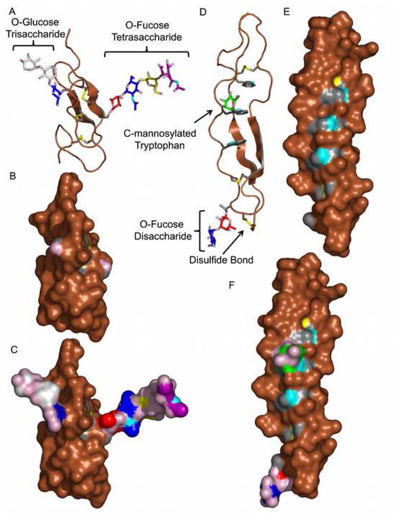
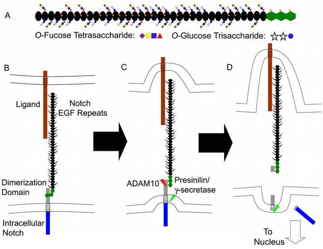
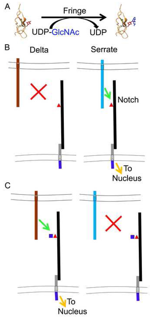
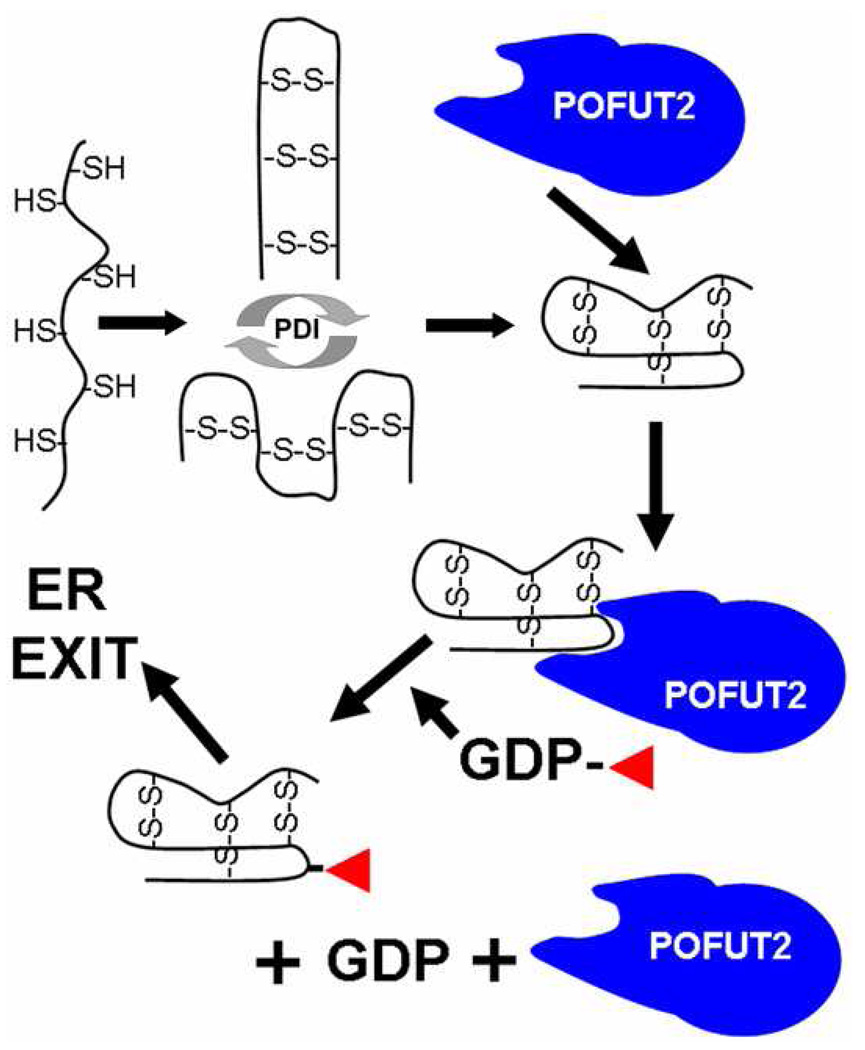
In the last two decades, our knowledge of the role of glycans in development and signal transduction has expanded enormously. While most work has focused on the importance of N-linked or mucin-type O-linked glycosylation, recent work has highlighted the importance of several more unusual forms of glycosylation that are the focus of this review. In particular, the ability of O-fucose glycans on the epidermal growth factor-like (EGF) repeats of Notch to modulate signaling places glycosylation alongside phosphorylation as a means to modulate protein-protein interactions and their resultant downstream signals. The recent discovery that O-glucose modification of Notch EGF repeats is also required for Notch function has further expanded the range of glycosylation events capable of modulating Notch signaling. The prominent role of Notch during development and in later cell-fate decisions underscores the importance of these modifications in human biology. The role of glycans in intercellular signaling events is only beginning to be understood and appears ready to expand into new areas with the discovery that thrombospondin type 1 repeats are also modified with O-fucose glycans. Finally, a rare form of glycosylation called C-mannosylation modifies tryptophans in some signaling competent molecules and may be a further layer of complexity in the field. We will review each of these areas focusing on the glycan structures produced, the consequence of their presence, and the enzymes responsible.


Figures

Figure 1
Figure 1. O-Glycan Containing Epidermal Growth Factor-like and Thrombospondin Type 1 Repeats
A) Cartoon representation of fully glycosylated Factor IX EGF repeat containing both an O-fucose tetrasaccharide (Sia-α2,3/6-Gal-β1,4-GlcNAc-β1,3-Fuc) on the right and an O-glucose trisaccharide (Xyl-α1,3-Xyl-α1,3-Glc ) on the left. The O-fucose tetrasaccharide containing EGF repeat was described previously (pdb accession 1EDM) (Luo, 2007). The O-glucose trisaccharide was modeled by taking a solution structure model from the Sweet website (http://www.glycosciences.de/modeling/sweet2/doc/index.php) and adding the modification to the appropriate serine. The serine side-chain was then cycled through known rotamer conformations and the position judged most appropriate by eye was chosen. B) Surface representation of panel A without glycosylation. C) Surface representation of panel A. D) Cartoon representation of a fully glycosylated TSR containing a Glc-β1,3-Fuc disaccharide and a mannosylated tryptophan. The disaccharide was modeled by taking a Glc-β1,3-Fuc solution structure model from the Sweet website, and aligning the fucose from the disaccharide with the fucose of TSR3 from the structure of the Thrombospondin 1 TSRs (pdb accession 1LSL) (Tan et al., 2002). The mannose on the tryptophan was modeled by superimposing the planar ring of 4-nitrophenyl-α-D-mannose with the tryptophan ring, removing the oxygen on C1 of the mannose, and placing the mannose at an approximated carbon bond length from the appropriate indole carbon. E) Surface representation of panel D without glycosylation. F) Surface representation of panel D. All panels are at the same scale. The carbons of sugar residues are colored according to the Consortium for Functional Glycomics (http://www.functionalglycomics.org) recommended nomenclature. Fucose is red, glucose and N-acetylglucosamine are blue, galactose is yellow, xylose is white, sialic acid is purple, and mannose is green. Oxygens are colored pink, nitrogen is cyan, carbons of amino acid side-chains involved in disulfide bonding or glycosylation are grey, and the sulfur of cysteines are colored yellow. All other atoms are brown. All pictures produced using PyMol (Delano, 2002).
Figure 2
Figure 2. Model of Notch Receptor Activation
A) Diagram of human Notch1 ECD. EGF repeats are indicated with black ovals, and Notch/Lin12 repeats as green hexagons. The glycans on Notch are indicated with the suggested shapes and colors of the Consortium for Functional Glycomics (http://www.functionalglycomics.org). Calcium ions are shown as orange spheres associated with EGF repeats and the Notch/Lin12 repeats. The glycan modifications and calcium binding EGF repeat pattern is based on human Notch 1. The diagram indicates a maximal level of glycosylation at all predicted sites. Mass spectral analysis suggests that this is not the case (Haltiwanger lab unpublished results, Nita-Lazar and Haltiwanger, 2006a, Nita-Lazar and Haltiwanger, 2006b, Xu et al, 2007), but that it varies depending on species, Notch subtype, and cell type. The in vivo glycosylation pattern could involve monosaccharide O-fucose or O-glucose on some repeats. Note that consecutive EGF repeats containing calcium binding sites are predicted to be rotated approximately 120° relative to one another (Hambleton et al., 2004). Thus the glycans are drawn pointing up or down (switching sides) on neighboring EGF repeats to reflect this. B–D) Notch activation. B) A simplified Delta ligand is shown on an adjacent cell in brown. The Delta ligand interacts with the Notch receptor on a neighboring cell. Note that the Lin12/Notch repeats interact non-covalently with the transmembrane/intracellular portion of Notch. The transmembrane portion of Notch is in grey, and the intracellular Notch is shown in simplified fashion in blue. C) Subsequent to Delta/Notch interaction, Delta begins to be endocytosed by the sending cell. This causes a conformational change in the Notch/Lin12 repeats exposing the cleavage site. ADAM10, which catalyzes the cleavage, is shown as a red lightning bolt. D) After the extracellular cleavage, the presenilin/γ-secretase complex (green lightening bolt) catalyzes the intracellular cleavage, releasing the intracellular domain from the membrane. The Notch intracellular domain transits to the nucleus to transactivate downstream targets.
Figure 3
Figure 3. Fringe Modulation of Notch Signaling
A. Diagram of the chemical reaction catalyzed by Fringe. The EGF repeat is drawn as described in Figure 1. The fucose is shown in red sticks, and the GlcNAc is shown in blue sticks. B. Notch activity without Fringe. Without Fringe, Notch is modified with O-fucose monosaccharide. Under these conditions, Serrate can activate Notch, but Delta cannot. Notch is drawn with a single O-fucose monosaccharide (red triangle) for simplicity. C. Notch activity with Fringe. With Fringe, Notch is modified with O-fucose disaccharide. Under these conditions, Delta can activate Notch, but Serrate cannot. As in panel B, Notch is drawn with a single disaccharide (GlcNAc, blue square; fucose, red triangle) for simplicity. The diagrams in panels A and B are drawn based on what is known about Drosophila Notch and Fringe.
Figure 4
Figure 4. Model of the Effects of Pofut2 on TSR Folding in the Endoplasmic Reticulum
As a protein containing TSR sequences is co-translationally translocated into the lumen of the ER, the cysteines emerge unpaired. For simplicity, the unpaired sulfhydryls of six cysteines from a single TSR are shown, although these typically occur in tandem repeats (e.g. ADAMTS13 has 8 TSRs (Ricketts et al., 2007)). Soon after entering the lumen, the TSR will enter a folding cycle catalyzed by chaperones and an oxidoreductase such as protein disulfide isomerase (PDI). A number of folding intermediates could result, although only one is properly folded. Since Pofut2 is ER localized and specifically recognizes properly folded TSRs (Luo et al., 2006a, Luo et al, 2006b), as soon as a properly folded TSR is formed, Pofut2 should bind. We have previously presented data that O-fucosylation of TSRs and EGF repeats occurs in the ER, suggesting the presence of GDP-fucose in the ER (Sturla et al., 2003). Thus, the TSR should be O-fucosylated at this point. O-Fucosylation may occur co-translationally, although definitive evidence for this does not yet exist. The O-fucosylated protein would then be ready for ER exit. Defects at any point in this process could affect the efficiency of ER exit. For instance mutation of O-fucosylation sites on the TSRs, elimination of Pofut2 by siRNA, or elimination of GDP-fucose in Lec13 cells, would all eliminate or reduce O-fucosylation. Each of these appears to affect secretion of ADAMTS13 or ADAMTS-like1 (Ricketts et al., 2007, Wang et al., 2007) which may be a result of reduced ER exit.

Similar articles

Cited by

References

    1. Acar M, Jafar-Nejad H, Takeuchi H, Rajan A, Ibrani D, Rana NA, et al. Rumi is a CAP10 domain glycosyltransferase that modifies Notch and is required for Notch signaling. Cell. 2008;132:247–258. - PMC - PubMed
    1. Adams JC, Lawler J. The thrombospondins. Int J Biochem Cell Biol. 2004;36:961–968. - PMC - PubMed
    1. Adams JC, Tucker RP. The thrombospondin type 1 repeat (TSR) superfamily: diverse proteins with related roles in neuronal development. Develop. Dynamics. 2000;218:280–299. - PubMed
    1. Andrade RP, Palmeirim I, Bajanca F. Molecular clocks underlying vertebrate embryo segmentation: A 10-year-old hairy-go-round. Birth Defects Res C Embryo Today. 2007;81:65–83. - PubMed
    1. Aoki K, Porterfield M, Lee SS, Dong B, Nguyen K, McGlamry KH, et al. The diversity of O-linked glycans expressed during Drosophila melanogaster development reflects stage-and tissue-specific requirements for cell signaling. J Biol Chem. 2008 (In Press) - PMC - PubMed

Publication types

LinkOut - more resources