. 2009 Jan 2;284(1):276-283.
doi: 10.1074/jbc.M805061200. Epub 2008 Oct 24.

Structure and proposed activity of a member of the VapBC family of toxin-antitoxin systems. VapBC-5 from Mycobacterium tuberculosis

Affiliations

Structure and proposed activity of a member of the VapBC family of toxin-antitoxin systems. VapBC-5 from Mycobacterium tuberculosis

Linda Miallau et al. J Biol Chem. .

Abstract

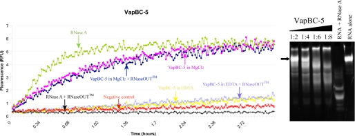
In prokaryotes, cognate toxin-antitoxin pairs have long been known, but no three-dimensional structure has been available for any given complex from Mycobacterium tuberculosis. Here we report the crystal structure and activity of a member of the VapBC family of complexes from M. tuberculosis. The toxin VapC-5 is a compact, 150 residues, two domain alpha/beta protein. Bent around the toxin is the VapB-5 antitoxin, a 33-residue alpha-helix. Assays suggest that the toxin is an Mg-enabled endoribonuclease, inhibited by the antitoxin. The lack of DNase activity is consistent with earlier suggestions that the complex represses its own operon. Furthermore, analysis of the interactions in the binding of the antitoxin to the toxin suggest that exquisite control is required to protect the bacteria cell from toxic VapC-5.


Figures

FIGURE 1.
FIGURE 1.
Sequence and structure of M. tuberculosis VapBC-5. A, ribbon diagram of the VapBC-5 complex (β-strands are cyan, α-helices are magenta for VapC-5 and green for VapB-5). Dotted lines represent residues that are not visible in the density. B, surface representation of VapC-5 showing negative electrostatic potential in red and positive in blue. VapB-5 is shown as a ribbon diagram with amino acid side chains shown in stick representation. Black arrows designate the cavities that shelter the active site residues as well as residues that may be involved in the binding of Mg2+ ions. C, amino acid sequence of M. tuberculosis VapC-5 on top and VapB-5 at the bottom with their secondary structure elements assigned and colored according to the ribbon diagram. Residues in lowercase are not seen in the structure. Putative catalytic residues are marked with a red star and Arg-102 with a blue star.
FIGURE 2.
FIGURE 2.
The putative active site of M. tuberculosis VapC-5. The main chain atoms are represented as a ribbon diagram and colored as follows: VapB-5 is shown in green, VapC-5 α-helices are shown in magenta, β-strands in cyan and loops in gray. Residues involved in the active site formation are shown as green sticks for VapB-5 and yellow for VapC-5. For both proteins, N and O atoms are blue and red, respectively. The four acidic catalytic residues are in bold font. Water molecules are shown as cyan balls. Potential hydrogen bonds are depicted as cyan-dashed lines. For the purpose of clarity, only selected water molecules and hydrogen bonds are shown.
FIGURE 3.
FIGURE 3.
Superposition of the structure of M. tuberculosis VapBC-5 with its structural homologues. In all figures, M. tuberculosis VapB-5 is shown in green, M. tuberculosis VapC-5 is in magenta, Ngo FitA (34) is in orange, Ngo FitB (34) is in red, and Pae VapC (14) is in blue. A, ribbon diagram of the superimposed M. tuberculosis VapBC-5 and Pae VapC, which shows that the structure between these two homologues is conserved except for helix α-2 in VapBC-5 that is shifted. B, top view of the zoomed region showing the large displacement of M. tuberculosis VapC-5 helix α-2 compared with the corresponding helix in Pae VapC-5. The disorganized loop linkingα-1 toα-2 is represented as red broken lines. C, ribbon diagram of the superimposed structures. The structure of the toxins in these different structural homologues is conserved whereas their respective cognates differ. D, zoom of the acidic cavity of the superimposed structures. It shows that Arg-112 from M. tuberculosis VapC-5 and Arg-68 from Ngo FitA both form hydrogen bonds with a residue that belongs to the active site and thus Arg-112 could play an indirect role in the mechanism of inhibition of the toxin. Residues from M. tuberculosis VapB-5 are shown as green sticks, those from M. tuberculosis VapC-5 are shown in yellow, Arg-68 from Ngo FitA is in orange, and active site residues from Ngo FitB are shown as red sticks. E, stereoview of the superposition of the putative active site residues of M. tuberculosis VapC-5 with the active site residues and magnesium ions of endo and exonuclease FEN-1 (31). Residues from M. tuberculosis VapC-5 are shown as yellow sticks, and active site residues and magnesium ions from endo and exonuclease FEN-1 are shown as gray sticks and green spheres, respectively. The conservation of most of the residues that bind the magnesium ions suggests that M. tuberculosis VapC-5 catalytic mechanism could involve the two metal ions as suggested by the mechanism of FEN-1 nuclease.
FIGURE 4.
FIGURE 4.
In vitro ribonuclease activity of M. tuberculosis VapBC-5. A, fluorescence measurements as a function of time. A fluorescent substrate is incubated with VapBC-5 in different conditions. Fluorescence is measured when the substrate is cleaved which indicates the presence of ribonuclease activity. It clearly shows that VapC-5 activity is dependent on the presence of magnesium as shown by the magenta and navy blue curves. B, nuclease assay: polyacrylamide/urea denaturing gel showing from left to right:2 μm RNA stained by Sybr Green II with increasing concentration of VapBC-5 in MgCl2. The RNA was incubated for 5 h at 37 °C with 4, 8, 12, 16 μm VapBC-5, respectively, and the subsequent lanes shows the RNA incubated with RNase A as a positive control and the RNA alone. The black arrow points to the intact RNA. Degradation products appear (smears) when the RNA is incubated with VapBC-5.

References

    1. Ogura, T., and Hiraga, S. (1983) Proc. Natl. Acad. Sci. U. S. A. 80 4784-4788 - PMC - PubMed
    1. Gerdes, K., Rasmussen, P. B., and Molin, S. (1986) Proc. Natl. Acad. Sci. U. S. A. 83 3116-3120 - PMC - PubMed
    1. Magnuson, R. D. (2007) J. Bacteriol. 189 6089-6092 - PMC - PubMed
    1. Buts, L., Lah, J., Dao-Thi, M. H., Wyns, L., and Loris, R. (2005) Trends Biochem. Sci. 30 672-679 - PubMed
    1. Pandey, D. P., and Gerdes, K. (2005) Nucleic Acids Res. 33 966-976 - PMC - PubMed

Publication types

MeSH terms

Associated data

LinkOut - more resources