Comment
. 2008 Oct;20(10):2546-9; author reply 2549-51.
doi: 10.1105/tpc.108.062299. Epub 2008 Oct 24.

A note on three-dimensional models of higher-plant thylakoid networks

Comment

A note on three-dimensional models of higher-plant thylakoid networks

Vlad Brumfeld et al. Plant Cell. 2008 Oct.
No abstract available


Figures

Figure 1.
Figure 1.
Reconstructed Structures of Granum-Stroma Assemblies. Tomographic data were obtained from cryofixed, freeze-substituted, dark-adapted lettuce leaf preparations. (A) The granum layers are contiguous with the stroma membranes that bifurcate (arrows) at the granum-stroma interface. Internal connections between adjacent layers are indicated by arrowheads. (B) To better visualize the internal connections between neighboring layers in the granum (arrowhead), the structure shown in (A) was enlarged, clipped, and rotated by ∼40°, and the upper layer of the stack has been removed. In both panels, the structure was expanded along the z axis to provide a clear view of the interior of the granum. (C) and (D) An ensemble of two grana interconnected by multiple stroma lamellae. The structure shown in (D) is a rotated view of the one shown in (C), with four of the lamellae removed. The stroma lamellae form wide, slightly undulating sheets that run parallel to each other and intersect the grana (surface-rendered gray objects) at an angle that is roughly perpendicular to the axis of the granum cylinder. The arrows shown in (C) define the plane of the layers inside the granum. All figures were adapted from Shimoni et al. (2005).
Figure 2.
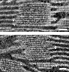
Figure 2.
Forks or Bifurcations Are Not Convoluted Slits. Shown are 2.6-nm-thick slices cut through the tomographic volume. Bifurcations of stroma lamellae into granum bodies are indicated by arrowheads. Bar = 65 nm.

Comment on

References

    1. Brangeon, J., and Mustárdy, L. (1979). Ontogenetic assembly of intra-chloroplastic lamellae viewed in 3-dimension. Biol Cell. 36 71–80.
    1. Heslop-Harrison, J. (1963). Structure and morphogenesis of lamellar systems in grana-containing chloroplasts. Planta 60 243–260.
    1. Hodge, A.J., McLean, J.D., and Mercer, F.V. (1955). Ultrastructure of the lamellae and grana in the chloroplasts of Zea mays. J. Biophys. Biochem. Cytol. 1 605–614. - PMC - PubMed
    1. Lučić, V., Förster, F., and Baumeister, W. (2005). Structural studies by electron tomography: From cells to molecules. Annu. Rev. Biochem. 74 833–865. - PubMed
    1. Mastronarde, D.N., Ladinsky, M.S., and McIntosh, J.R. (1997). Super-thin serial sectioning for high-resolution 3-D reconstruction of cellular structures. Microsc. Microanal. 3 221–222.

LinkOut - more resources