. 2008 Nov 4;105(44):17103-8.
doi: 10.1073/pnas.0802696105. Epub 2008 Oct 24.

Disruption of the NHR4 domain structure in AML1-ETO abrogates SON binding and promotes leukemogenesis

Affiliations

Disruption of the NHR4 domain structure in AML1-ETO abrogates SON binding and promotes leukemogenesis

Eun-Young Ahn et al. Proc Natl Acad Sci U S A. .

Abstract

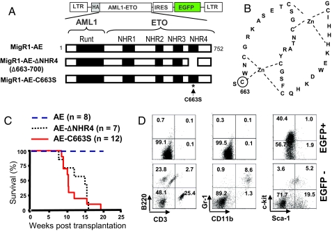
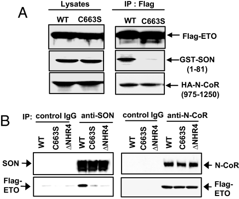
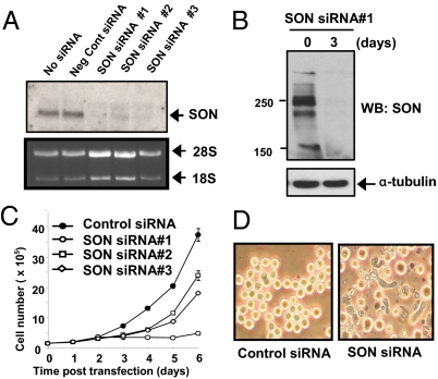
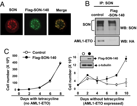
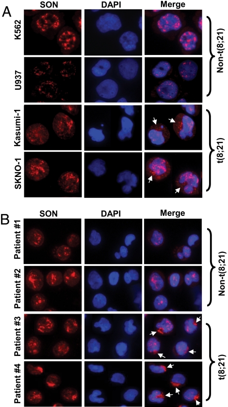
AML1-ETO is generated from t(8;21)(q22;q22), which is a common form of chromosomal translocation associated with development of acute myeloid leukemia (AML). Although full-length AML1-ETO alone fails to promote leukemia because of its detrimental effects on cell proliferation, an alternatively spliced isoform, AML1-ETO9a, without its C-terminal NHR3/NHR4 domains, strongly induces leukemia. However, full-length AML1-ETO is a major form of fusion product in many t(8;21) AML patients, suggesting additional molecular mechanisms of t(8;21)-related leukemogenesis. Here, we report that disruption of the zinc-chelating structure in the NHR4 domain of AML1-ETO by replacing only one critical amino acid leads to rapid onset of leukemia, demonstrating that the NHR4 domain with the intact structure generates inhibitory effects on leukemogenesis. Furthermore, we identified SON, a DNA/RNA-binding domain containing protein, as a novel NHR4-interacting protein. Knock-down of SON by siRNA resulted in significant growth arrest, and disruption of the interaction between AML1-ETO and endogenous SON rescued cells from AML1-ETO-induced growth arrest, suggesting that SON is an indispensable factor for cell growth, and AML1-ETO binding to SON may trigger signals inhibiting leukemogenesis. In t(8;21) AML patient-derived primary leukemic cells and cell lines, abnormal cytoplasmic localization of SON was detected, which may keep cells proliferating in the presence of full-length AML1-ETO. These results uncovered the crucial role of the NHR4 domain in determination of cellular fate during AML1-ETO-associated leukemogenesis.


Conflict of interest statement

The authors declare no conflict of interest.

Figures

Fig. 1.
Fig. 1.
Deletion of the NHR4 domain or a point mutation at the Zn-chelating residue of the NHR4 domain of AML1-ETO causes rapid onset of leukemia. (A) Schematic representation of AML1-ETO fusion protein and the retroviral construct MigR1. Three constructs were used in the retroviral infection and transplantation experiments: full-length AML-ETO (MigR1-AE), NHR4 domain-deleted AML1-ETO (MigR1-AE-ΔNHR4), and AML1-ETO with serine substitution of cysteine at a.a. 663 (MigR1-AE-C663S). (B) Schematic representation of the Zn-chelating structures in NHR4 of AML1-ETO. One of the Zn-chelating residues, cysteine residue at position 663 (marked with a circle), was chosen for a point mutation that would disrupt the Zn-chelating structure of NHR4 domain. (C) Kaplan-Meier survival curves of MF-1 mice transplanted with fetal liver cells retrovirally-transduced with MigR1-AE, MigR1-AE-ΔNHR4, and MigR1-AE-C663S. (D) Flow-cytometric analysis of lineage markers expressed in EGFP-positive (EGFP+) and EGFP-negative (EGFP-) populations of peripheral blood from the leukemic mice with MigR1-AE-ΔNHR4. The numbers in each quadrant represent the percentage of cells.
Fig. 2.
Fig. 2.
Identification of SON as an NHR4 domain-interacting protein. (A) Schematic representation of three baits used for yeast two hybrid screening and their interaction with the N-terminal 102 a.a. of SON in yeast; full-length wild-type ETO (ETO-wt), full-length ETO with a point mutation at the NHR4 domain (ETO-C663S), and the C-terminal region of ETO (NHR3-NHR4). Binding activities were determined (-, +, and ++) by expression of β-galactosidase reporter gene and the growth of yeast colonies in URA- media. (B) Interaction of the SON N-terminal fragments with ETO in mammalian cells. The N-terminal region of mouse SON encoding a.a 1–25, 1–81, or 1–102 were fused with GST, co-expressed with Flag-tagged ETO in 293T cells, and GST pull-down complexes were immunoblotted with GST antibody or Flag antibody. Note that the GST control shows a similar molecular weight to GST-SON-1–25, because the translational termination site for GST alone is located further downstream of the multiple cloning sites, resulting in the additional 20 a.a. to the C terminus of GST. (C) Interaction of endogenous SON and AML1-ETO in t(8;21) leukemia patient-derived cell line, SKNO-1. The SON antibody pulled down AML1-ETO in SKNO-1 cell lysates. SON was detected as several bands, although its calculated molecular weight is ≈267 kDa. A band marked with an asterisk is nonspecific. (D) Colocalization of SON and AML1-ETO. HeLa cells were transfected with HA-AML1-ETO or Flag-ETO and immunostained with HA- or Flag-antibody and SON antibody followed by confocal microscopy.
Fig. 3.
Fig. 3.
Deletion of NHR4 or C663S mutation abolishes SON interaction but not N-CoR interaction. (A) Effects of the C663S mutation on interactions with the SON N-terminal fragment and the N-CoR fragment containing the RIII domain. GST-tagged SON (1–81 a.a.) and HA-tagged N-CoR (975–1250 a.a.) were transfected into 293T cells with either Flag-tagged wild-type ETO (WT) or ETO with C663S mutation, and immunoprecipited with Flag antibody. (B) Effects of NHR4 deletion or C663S mutation on interactions with full-length SON and N-CoR. SON antibody or N-CoR antibody was used for immunoprecipitation to pull down endogenous SON or N-CoR, and immunoblotted to detect Flag-tagged ETO; wild-type ETO (WT), ETO with a C663S mutation (C663S), and NHR4-deleted ETO (ΔNHR4).
Fig. 4.
Fig. 4.
SON siRNA completely knocks down endogenous SON and induces growth arrest. (A) Knock-down of SON by SON siRNAs in 293T cells confirmed by Northern blotting. (B) Knock-down of SON confirmed by Western blotting. K562 cells were transfected with SON siRNA no. 1. Whole cell lysates were prepared after 3 days and immunoblotted with SON antibody. (C) SON siRNA induces growth arrest. K562 cells were transfected with 1.3 μM negative control siRNA or three different SON siRNAs by nucleofection in 100 μl solution, and cells were counted every day. The graph is representative of four experiments. (D) Morphological changes in K562 cells 3 days after SON siRNA (#1) transfection.
Fig. 5.
Fig. 5.
Expression of SON N-terminal fragment inhibits AML1-ETO binding to endogenous SON, and rescues cells from AML1-ETO-induced growth arrest. (A) The SON N-terminal fragment encoding a.a. 1–140 (Flag-SON-140) colocalizes with endogenous SON. K562 cells expressing MigR1-Flag-SON-140 were immunostained with SON antibody (recognizing C terminus of SON) for endogenous SON, and with Flag antibody for the SON-140 fragment. (B) Expression of the SON-140 fragment interferes with the interaction between endogenous SON and AML1-ETO. K562 cells stably expressing control vector or Flag-SON-140 were transfected with HA-tagged AML1-ETO, and immunoprecipitation was performed with SON antibody, and the immunocomplexes were subjected to Western blotting with the indicated antibodies. (C) Expression of the SON-140 fragment alone does not affect cell growth and rescues cells from AML1-ETO-induced growth arrest. U937T cells with inducible AML1-ETO expression were infected with control vector or Flag-SON-140. The GFP-positive populations were sorted, cultured with or without tetracycline, and cells were counted every other day. Western blot shows expression of Flag-tagged SON-140 in sorted cells. Growth curve shown represents four independent experiments. Each circle and error bar represents mean ± SD, from the triplicate cell culture.
Fig. 6.
Fig. 6.
Abnormal cytoplasmic localization of SON in t(8;21)-positive leukemic cells. (A) Two leukemic cell lines without t(8;21), K562 and U937, and two t(8;21) leukemic cell lines, Kasumi- 1 and SKNO-1, were stained with SON antibody and DAPI (for DNA). (B) Blood cells from four different AML patients were stained with SON antibody and DAPI, and analyzed by fluorescence microscopy. Patient 1, FAB M2 subtype, non-t(8;21); patient 2, FAB M4 subtype, non-t(8;21); patients 3 and 4, FAB M2 subtype, t(8;21) -positive. Arrows indicate cytoplasm-localized SON. Pictures are of representative features of each cell line and patient samples from >1000 cells analyzed by fluorescence microscopy.

References

    1. Rowley JD. Chromosome translocations: Dangerous liaisons revisited. Nat Rev Cancer. 2001;1:245–250. - PubMed
    1. Langabeer SE, et al. Incidence of AML1/ETO fusion transcripts in patients entered into the MRC AML trials. MRC Adult Leukaemia Working Party. Br J Haematol. 1997;99:925–928. - PubMed
    1. Nucifora G, Rowley JD. AML1 and the 8;21 and 3;21 translocations in acute and chronic myeloid leukemia. Blood. 1995;86:1–14. - PubMed
    1. Rege K, et al. Disease features in acute myeloid leukemia with t(8;21)(q22;q22). Influence of age, secondary karyotype abnormalities, CD19 status, and extramedullary leukemia on survival. Leuk Lymphoma. 2000;40:67–77. - PubMed
    1. Rowe D, et al. Cytogenetically cryptic AML1-ETO and CBF beta-MYH11 gene rearrangements: Incidence in 412 cases of acute myeloid leukaemia. Br J Haematol. 2000;111:1051–1056. - PubMed

Publication types

MeSH terms

Substances