. 2008 Nov 4;105(44):17127-32.
doi: 10.1073/pnas.0804387105. Epub 2008 Oct 24.

Predataxis behavior in Myxococcus xanthus

Affiliations

Predataxis behavior in Myxococcus xanthus

James E Berleman et al. Proc Natl Acad Sci U S A. .

Abstract

Spatial organization of cells is important for both multicellular development and tactic responses to a changing environment. We find that the social bacterium, Myxococcus xanthus utilizes a chemotaxis (Che)-like pathway to regulate multicellular rippling during predation of other microbial species. Tracking of GFP-labeled cells indicates directed movement of M. xanthus cells during the formation of rippling wave structures. Quantitative analysis of rippling indicates that ripple wavelength is adaptable and dependent on prey cell availability. Methylation of the receptor, FrzCD is required for this adaptation: a frzF methyltransferase mutant is unable to construct ripples, whereas a frzG methylesterase mutant forms numerous, tightly packed ripples. Both the frzF and frzG mutant strains are defective in directing cell movement through prey colonies. These data indicate that the transition to an organized multicellular state during predation in M. xanthus relies on the tactic behavior of individual cells, mediated by a Che-like signal transduction pathway.


Conflict of interest statement

The authors declare no conflict of interest.

Figures
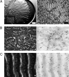
Fig. 1.
Fig. 1.
Multicellular reorganization during predation. (A) Stereo microscopy (Left) of M. xanthus strain DZ2 penetrating an E. coli prey colony and inducing rippling behavior. The image is centered on the prey colony, where a majority of the prey cells have been lysed and are no longer visible, but a fraction of the remaining E. coli colony is visible as a dark crescent on the Right edge. The open box denotes the area of the phase contrast image (Right) which is centered on the near edge of the prey colony where the transition from nonrippling to rippling behavior can be observed. (White scale bar, 1 mm; Black scale bar, 100 μm.) (B) Phase contrast (Left) and fluorescence microscopy (Right) of DZ2 mixed in a 50:1 ratio with GFP-labeled DZ10547, showing the characteristic mesh pattern of M. xanthus gliding behavior in the absence of prey. (C) Within the prey colony, the rippling pattern is stably maintained, although individual cells change position. (White scale bars in b and c, 10 μm.)
Fig. 2.
Fig. 2.
Tracking movement of M. xanthus cells. Tracking of GFP-labeled cells in (A) the absence of prey and (B) the presence of E. coli prey. Experimental setup is the same as in Fig. 1. Thirty cell tracks are shown for each condition. Cell movement over time is represented by the change in color of each track, with the beginning of the track labeled in dark blue and the end in dark red. Vector displacements of 100 cells for each condition are shown on the Right. The direction of migration through the E. coli prey colony is from Left to Right. (C) Distribution of cells was monitored at 2-min intervals by measuring fluorescence intensity across the entire field of view. The dashed line indicates the position of the wave crests at 0 min, and the arrows show the general direction of cell movement in the given area. The wave structure dissipates as M. xanthus cells leave the initial wave aggregates, but a similar pattern of aggregation emerges, shifted by ½ wavelength, at 8 min.
Fig. 3.
Fig. 3.
Quantitative analysis of rippling pattern dynamics. (A) Rippling wavelength as a function of incubation time during predation. A 1-μl suspension of 106 total M. xanthus strain DZ2 cells pipetted onto CFL media adjacent to a 3-μl colony containing 5 × 10⁷ cells of E. coli β2155 prey. The dashed line indicates times when quantifiable rippling was not observed. (B) Rippling wavelength at 40 h changes as a function of E. coli β2155 prey cell density (2 × 10⁶ to 6 × 10⁷ total cells) in M. xanthus strain DZ2 (diamonds), the wavelength is constitutively short in ΔfrzG (open sqaures), and no rippling is observed in ΔfrzF (−). (C) Rippling wavelength shows no change as a function of M. xanthus strain DZ2 cell density at 40 h (2 × 10⁵ to 4 × 10⁷ total cells) incubated with 5 × 10⁷ cells of E. coli β2155 prey. (D) Rippling induction time changes as a function of M. xanthus strain DZ2 cell density (6 × 10² to 1 × 10⁷ total cells) incubated with 5 × 10⁷ cells of E. coli β2155 prey.
Fig. 4.
Fig. 4.
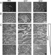
Cooperation analysis in adaptation mutants. A 10-μl aliquot containing 107 M. xanthus strain DZ2 cells was pipetted on CFL agar in the absence of prey. Stereo and phase microscopy images were captured at 72 h at (A) the colony edge at which group gliding behavior can be observed and (B) the colony center where fruiting body aggregation is observed. A ΔfrzF mutant is defective in forming gliding groups and also forms unorganized “frizzy” aggregates. ΔfrzG displays a phenotype with only subtle differences from the wild type in the absence of prey. (C) After 40 h in coculture with E. coli prey, wild-type cells modulate their rippling wavelength in response to the availability of prey. The ΔfrzF mutant does not form ripple structures. The ΔfrzG mutant forms numerous ripples at all prey cell densities in which rippling is induced. The direction of migration through E. coli prey is from Left to Right. (Black bar, 100 μm in a and c; White bar, 1 mm (b).)
Fig. 5.
Fig. 5.
Immunoblot analysis of the methylation state of the FrzCD receptor. Cells of DZ2 (wild type, lanes 1–3), ΔfrzF (cheR, lanes 4–6), and ΔfrzG (cheB, lanes 7–9) were harvested after 24-h growth in liquid CYE (lanes 1, 4, and 7) and plated on CFL media (108 total cells) either in the absence (lanes 2, 5, and 8) or presence of 109 E. coli prey cells (lanes 3, 6, and 9). Cells were harvested after 24 h at 32 °C and all samples were analyzed using SDS/PAGE to separate the methylated and demethylated forms of FrzCD, as described previously (26). frzCDc, a strain that expresses a truncated form of FrzCD, was used as a negative control (lane 10).
Fig. 6.
Fig. 6.
Tactic assay of predatory behavior. A 1-μl aliquot containing 106 M. xanthus cells was pipetted in the center of a 40 × 2.5 mm strip of E. coli β2155 containing a total of 4 × 10⁸ cells. Swarm expansion was measured in two axes; the x-axis contains prey cells (closed shapes), the y-axis does not (open shapes). The arrows indicate when rippling behavior begins. (A) Assay diagram, (B) M. xanthus strain DZ2, (C) ΔfrzG, and (D) ΔfrzF.

References

    1. King N. The unicellular ancestry of animal development. Dev Cell. 2004;7:313–325. - PubMed
    1. Aguilar C, Vlamakis H, Losick R, Kolter R. Thinking about Bacillus subtilis as a multicellular organism. Curr Opin Microbiol. 2007;10:638–643. - PMC - PubMed
    1. Camilli A, Bassler BL. Bacterial small-molecule signaling pathways. Science. 2006;311:1113–1116. - PMC - PubMed
    1. Zusman DR, Scott AE, Yang Z, Kirby JR. Chemosensory pathways, motility and development in Myxococcus xanthus. Nat Rev Microbiol. 2007;5:862–872. - PubMed
    1. Mignot T, Merlie JP, Jr, Zusman DR. Regulated pole-to-pole oscillations of a bacterial gliding motility protein. Science. 2005;310:855–857. - PubMed

Publication types

Substances