. 2008 Nov 11;105(45):17504-9.
doi: 10.1073/pnas.0804054105. Epub 2008 Oct 24.

Role of TGF-beta in proliferative vitreoretinal diseases and ROCK as a therapeutic target

Affiliations

Role of TGF-beta in proliferative vitreoretinal diseases and ROCK as a therapeutic target

Takeshi Kita et al. Proc Natl Acad Sci U S A. .

Abstract

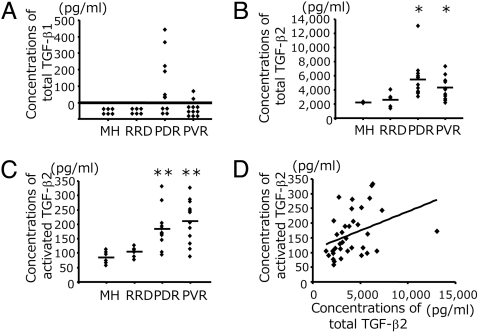
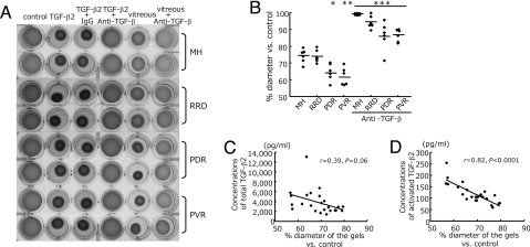
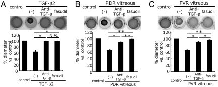
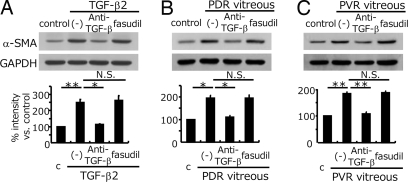
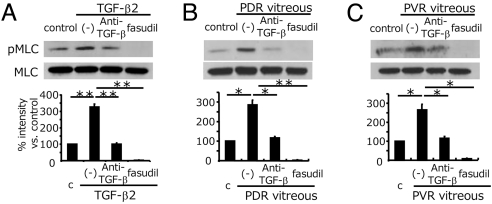
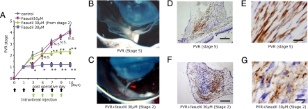
Cicatricial contraction of preretinal fibrous membrane is a cause of severe vision loss in proliferative vitreoretinal diseases such as proliferative diabetic retinopathy (PDR) and proliferative vitreoretinopathy (PVR). TGF-beta is overexpressed in the vitreous of patients with proliferative vitreoretinal diseases and is also detectable in the contractile membranes. Therefore, TGF-beta is presumed to contribute to the cicatricial contraction of the membranes, however, the underlying mechanisms and TGF-beta's importance among various other factors remain to be elucidated. Vitreous samples from PDR or PVR patients caused significantly larger contraction of hyalocyte-containing collagen gels, compared with nonproliferative controls. The contractile effect was strongly correlated with the vitreal concentration of activated TGF-beta2 (r = 0.82, P < 0.0001). PDR or PVR vitreous promoted expression of alpha-smooth muscle actin (alpha-SMA) and phosphorylation of myosin light chain (MLC), a downstream mediator of Rho-kinase (ROCK), both of which were dramatically but incompletely suppressed by TGF-beta blockade. In contrast, fasudil, a potent and selective ROCK inhibitor, almost completely blocked the vitreous-induced MLC phosphorylation and collagen gel contraction. Fasudil disrupted alpha-SMA organization, but it did not affect its vitreal expression. In vivo, fasudil significantly inhibited the progression of experimental PVR in rabbit eyes without affecting the viability of retinal cells by electroretinographic and histological analyses. These results elucidate the critical role of TGF-beta in mediating cicatricial contraction in proliferative vitreoretinal diseases. ROCK, a key downstream mediator of TGF-beta and other factors might become a unique therapeutic target in the treatment of proliferative vitreoretinal diseases.


Conflict of interest statement

The authors declare no conflict of interest.

Figures

Fig. 1.
Fig. 1.
Concentrations of total and activated TGF-β1 and -β2 protein in the vitreous. (A) Concentrations of total TGF-β1 in the vitreal samples were measured by ELISA (MH, n = 6; RRD, n = 6; PDR, n = 12; and PVR, n = 11). (B and C) Concentrations of total TGF-β2 (B) and activated TGF-β2 (C). (MH, n = 6; RRD, n = 6; PDR, n = 12; and PVR, n = 12). *: PDR vs. MH, P = 0.0007; PDR vs. RRD, P = 0.003; PVR vs. MH, P = 0.002; and PVR vs. RRD, P = 0.049. **: PDR vs. MH, P = 0.005; PDR vs. RRD, P = 0.015; PVR vs. MH, P = 0.002; and PVR vs. RRD, P = 0.005 (Mann-Whitney U-test). (D) Correlation between the concentrations of total and activated TGF-β2 (r = 0.37, P = 0.026).
Fig. 2.
Fig. 2.
Role of TGF-β in vitreous-induced collagen gel contraction. (A) Hyalocyte-containing collagen gels were exposed to control (DMEM), recombinant TGF-β2 (0.3 nM), or patient vitreous, with anti-TGF-β mAb (10 ng/ml) or control IgG (10 ng/ml) (n = 6 in each group). Two representative wells per condition, 3 days after stimulation, are shown. (B) The diameters of the gels treated with vitreous (lane 5 in A and vitreous with anti-TGF-β mAb lane 6 in A were measured and expressed as a percentage of the diameter of control (lane 1 in A). *, P = 0.01 vs. MH and P = 0.007 vs. RRD; **, P = 0.007 vs. MH and P = 0.004 vs. RRD; ***, P < 0.0001 vs. each disease without anti-TGF-β mAb. (C and D) The correlation of the diameters of the gels with concentration of total TGF-β2 in the vitreous (n = 24, r = 0.39, P = 0.06) (C) and with activated TGF-β2 (n = 24, r = 0.82, P < 0.0001) (D).
Fig. 3.
Fig. 3.
Suppressive effect of fasudil on PDR/PVR-induced collagen gel contraction. Hyalocyte-containing collagen gels were stimulated with recombinant TGF-β2 (A), PDR vitreous (B), or PVR vitreous (C) (n = 3, each group) with or without anti-TGF-β mAb or fasudil (20 μM) for 3 days. Thereafter, the gels were photographed and the diameter of the gels was measured and statistically analyzed. Representative results are shown. *, P < 0.05; **, P < 0.01; NS, not significant (paired t test).
Fig. 4.
Fig. 4.
Role of TGF-β in the enhanced vitreous-induced α-SMA expression. The gels from Fig. 3 were dissolved and the cells were isolated and lysed. α-SMA expressions were examined by Western blot analysis. Loaded cell lysates in A, B, and C were from the gels in Fig. 3 A, B, and C, respectively. Representative blots are shown. Signal intensities were quantified and expressed as percentages of the α-SMA/GAPDH ratio compared with control (treated with DMEM). *, P < 0.05; **, P < 0.01; NS, not significant (paired t test).
Fig. 5.
Fig. 5.
Impact of fasudil on vitreous-induced MLC phosphorylation. After pretreatment with or without anti-TGF-β mAb or fasudil, hyalocytes were stimulated with recombinant TGF-β2 (A), vitreous with PDR (B), or vitreous with PVR (C) for 24 h (n = 3, each). Western blot analysis was performed to detect phosphorylated MLC (pMLC). Lane-loading differences were normalized by MLC. Signal intensities were quantified and expressed as percentages of the pMLC/MLC ratio compared with control (treated with DMEM). *, P < 0.05; **, P < 0.01.
Fig. 6.
Fig. 6.
Experimental PVR in rabbit eyes. (A) Therapeutic potential of fasudil in reducing the progression of experimental PVR. PVR was classified into six stages (0–5). Rhombus, vehicle (n = 5); purple square, fasudil 10 μM (n = 5); trigone, fasudil 30 μM from stage 2 (n = 6); blue square, fasudil 30 μM (n = 5). *, P < 0.05; **, P < 0.01;, not significant vs. vehicle. (B and C) Tractional retinal detachment because of formation and cicatricial contraction of preretinal proliferative membrane was observed by stereomicroscopy in vehicle-treated eyes (stage 5 PVR). (D and F) In contrast, intravitreal membranes adhered to the retina without causing retinal detachment (arrowhead) in 30 μM fasudil-treated eyes with stage 2 PVR. Micrographs depict α-SMA expression (brown) in preretinal proliferative membrane with stage 5 PVR (D) and stage 2 PVR (F) by immunohistochemical analysis. (Scale bar, 200 μm.) (E and G) Magnified images of D and F, respectively. (Scale bar, 10 μm.)

References

    1. Fong DS, et al. American Diabetes Association: Diabetic retinopathy. Diabetes Care. 2003;26:S99–S102. - PubMed
    1. Pastor JC, de la Rua ER, Martin F. Proliferative vitreoretinopathy: Risk factors and pathology. Prog Retin Eye Res. 2002;21:127–144. - PubMed
    1. Kampik A, Kenyon KR, Michels RG, Green WR, de la Cruz ZC. Epiretinal and vitreous membranes: Comparative study of 56 cases. Arch Ophthalmol. 1981;99:1445–1454. - PubMed
    1. Jerdan JA, et al. Proliferative vitreoretinopathy membranes: An immunohistochemical study. Ophthalmology. 1989;96:801–810. - PubMed
    1. Salu P, Claeskens W, De Wilde A, Hijmans W, Wisse E. Light and electron microscopic studies of the rat hyalocyte after perfusion fixation. Ophthalmic Res. 1985;17:125–130. - PubMed

Publication types

MeSH terms