. 2008 Nov 4;105(44):17115-20.
doi: 10.1073/pnas.0805239105. Epub 2008 Oct 24.

Mouse and human phenotypes indicate a critical conserved role for ERK2 signaling in neural crest development

Affiliations

Mouse and human phenotypes indicate a critical conserved role for ERK2 signaling in neural crest development

Jason Newbern et al. Proc Natl Acad Sci U S A. .

Abstract

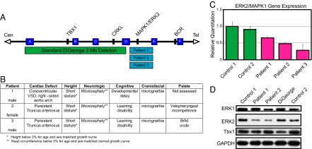
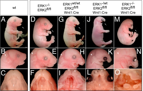
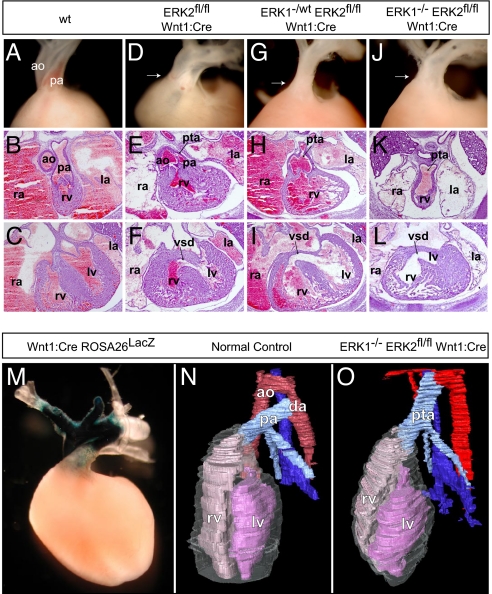
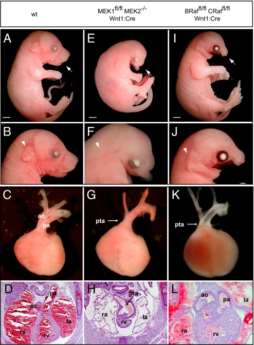
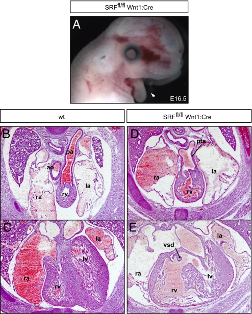
Disrupted ERK1/2 (MAPK3/MAPK1) MAPK signaling has been associated with several developmental syndromes in humans; however, mutations in ERK1 or ERK2 have not been described. We demonstrate haplo-insufficient ERK2 expression in patients with a novel approximately 1 Mb micro-deletion in distal 22q11.2, a region that includes ERK2. These patients exhibit conotruncal and craniofacial anomalies that arise from perturbation of neural crest development and exhibit defects comparable to the DiGeorge syndrome spectrum. Remarkably, these defects are replicated in mice by conditional inactivation of ERK2 in the developing neural crest. Inactivation of upstream elements of the ERK cascade (B-Raf and C-Raf, MEK1 and MEK2) or a downstream effector, the transcription factor serum response factor resulted in analogous developmental defects. Our findings demonstrate that mammalian neural crest development is critically dependent on a RAF/MEK/ERK/serum response factor signaling pathway and suggest that the craniofacial and cardiac outflow tract defects observed in patients with a distal 22q11.2 micro-deletion are explained by deficiencies in neural crest autonomous ERK2 signaling.


Conflict of interest statement

The authors declare no conflict of interest.

Figures

Fig. 1.
Fig. 1.
ERK2/MAPK1 protein and mRNA levels are decreased in patients with distal 22q11.2 deletions. (A) Deletions of chromosome 22q11.2 and their endpoints. Chromosome-specific low copy repeats are designated A through F (not to scale). The distal deletions seen in the patients in this study occur in the 1-Mb interval between low copy repeats (D and E). Relative locations of relevant genes are given. (B) Clinical findings in patients with distal micro-deletions of chromosome 22q11.2. (C) Gene expression plot from TaqMan assay of ERK2/MAPK1 is shown compared with 18s rRNA endogenous control. Error bars represent a composite of 2 separate experiments run in triplicate. (D) Western blotting of lymphoblastoid samples revealed decreased ERK2, but not TBX1, in 2 different patients with distal 22q11.2 micro-deletions compared with normal controls. Samples from a patient with DGS show decreased TBX1, but no change in ERK2.
Fig. 2.
Fig. 2.
The development of craniofacial structures in mice is critically dependent on ERK2 expression in neural crest. In comparison to E17.5 WT embryos (A–C) ERK1−/− embryos exhibit no significant defects (D–F). E17.5 ERK2fl/fl Wnt1:Cre embryos exhibit deficits in maxilla formation and mandibular hypoplasia (G and H), cleft palate (I, arrow), and absence of the tongue. Interestingly, ERK1−/wt ERK2fl/fl Wnt1:Cre show more severe defects in craniofacial development (J–L). ERK1−/− ERK2fl/fl Wnt1:Cre embryos exhibited the most significant defects, including truncation of the maxilla (M and N), mandibular aplasia (M and N), absence of an external ear (arrowhead in N), overall decrease in crown–rump length, eye placement anomalies, and palatal defects (O, arrow). Scale bars, 2 mm.
Fig. 3.
Fig. 3.
Disruption of ERK1/2 signaling results in cardiac outflow tract defects. Compared with controls (A–C), ERK2fl/fl Wnt1:Cre embryos displayed variable penetrance of cardiac outflow defects, including double-outlet right ventricle (D), PTA (E), and VSDs (F). E16.5 cross-sections and dissected E17.5 embryos (atria dissected away) from ERK1−/wt ERK2fl/fl Wnt1:Cre (G–I) and ERK1−/− ERK2fl/fl Wnt1:Cre (J–L) embryos consistently exhibited PTA and VSDs (I and L). Whole-mount LacZ staining of Wnt1:Cre Rosa26LacZ hearts reveals the distribution of neural crest derivatives in the embryonic conotruncus (M). Three-dimensional reconstructions (N) of cross-sections from E16.5 control embryos show a normal heart with 2 separate vessels, the aorta (red) and pulmonary artery (light blue), connected distally by the ductus arteriosus. Cross-sectional reconstructions of ERK1−/− ERK2fl/fl Wnt1:Cre hearts further illustrate PTA in these embryos (O). (ao = aorta, pa = pulmonary artery, rv = right ventricle, lv = left ventricle, la = left atrium, ra = right atrium, da = ductus arteriosus.)
Fig. 4.
Fig. 4.
Inactivation of upstream elements of the MAPK cascade results in analogous defects in neural crest development. Compared with WT controls (A–D), mandibular hypoplasia and maxillary truncation is observed in MEK1fl/fl MEK2−/− Wnt1:Cre E17.5 embryos (E and F). Disruption of MEK1/2 signaling also resulted in decreased crown–rump length, eye placement anomalies, absence of the tongue, and a disruption of external ear development (E and F). PTA was consistently detected in E16.5 cross-sections and dissected E17.5 MEK1fl/fl MEK2−/− Wnt1:Cre embryos (G and H). The conditional inactivation of B-Raf/C-Raf did not alter embryonic size or external ear development, but mandibular and maxilla hypoplasia (I and J) was observed. Analysis of B-Raffl/fl C-Raffl/fl Wnt1:Cre embryos revealed partial penetrance of cardiac defects. An E17.5 embryo is displayed (K) that clearly demonstrates PTA whereas cross-sections from the E16.5 embryo (L) show mild conotruncal defects, namely double-outlet right ventricle. Scale bars, 2 mm.
Fig. 5.
Fig. 5.
Neural crest–specific inactivation of SRF disrupts mouse development. E16.5 SRFfl/fl Wnt1:Cre embryos exhibit fully penetrant mandibular hypoplasia (A, arrowhead). These embryos possess cardiac defects, including PTA (D) with VSDs (E). Scale bars, 2 mm.

References

    1. Pearson G, et al. Mitogen-activated protein (MAP) kinase pathways: Regulation and physiological functions. Endocr Rev. 2001;22:153–183. - PubMed
    1. Posern G, Treisman R. Actin' together: Serum response factor, its cofactors and the link to signal transduction. Trends Cell Biol. 2006;16:588–596. - PubMed
    1. Shaw PE, Saxton J. Ternary complex factors: Prime nuclear targets for mitogen-activated protein kinases. Int J Biochem Cell Biol. 2003;35:1210–1226. - PubMed
    1. Schubbert S, Bollag G, Shannon K. Deregulated Ras signaling in developmental disorders: New tricks for an old dog. Curr Opin Genet Dev. 2007;17:15–22. - PubMed
    1. Bentires-Alj M, Kontaridis MI, Neel BG. Stops along the RAS pathway in human genetic disease. Nat Med. 2006;12:283–285. - PubMed

Publication types

MeSH terms

Substances