. 2008 Nov;15(11):1176-1183.
doi: 10.1038/nsmb.1476. Epub 2008 Oct 26.

De novo DNA methylation promoted by G9a prevents reprogramming of embryonically silenced genes

Affiliations

De novo DNA methylation promoted by G9a prevents reprogramming of embryonically silenced genes

Silvina Epsztejn-Litman et al. Nat Struct Mol Biol. 2008 Nov.

Abstract

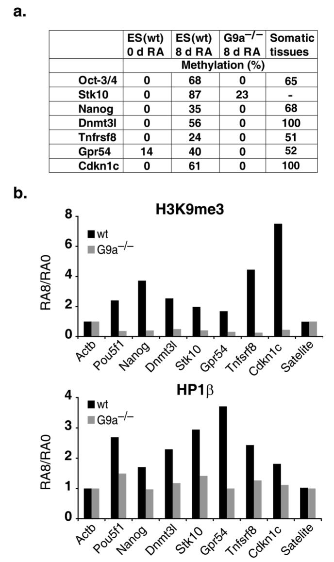
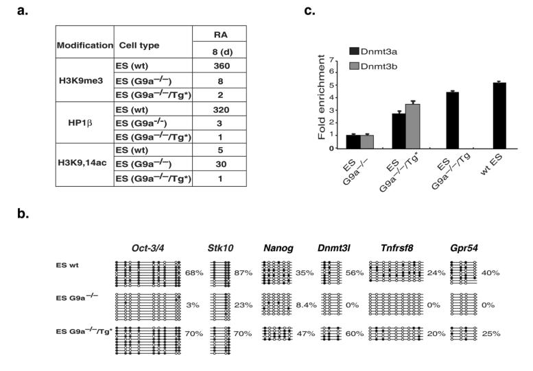
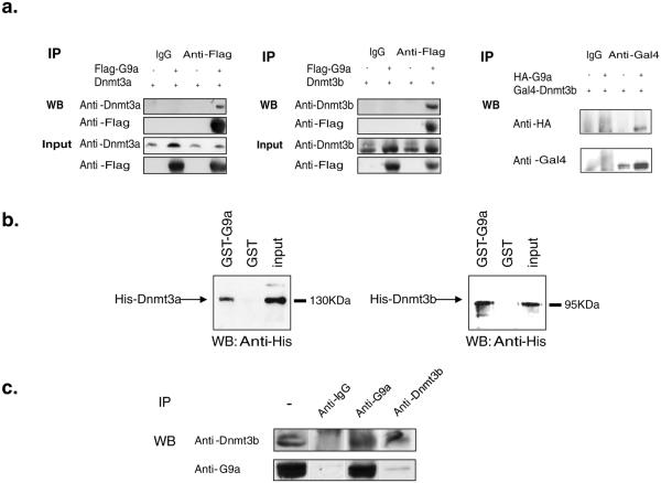
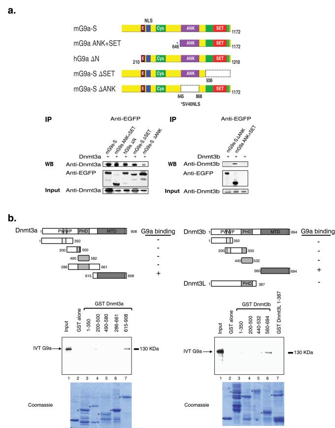
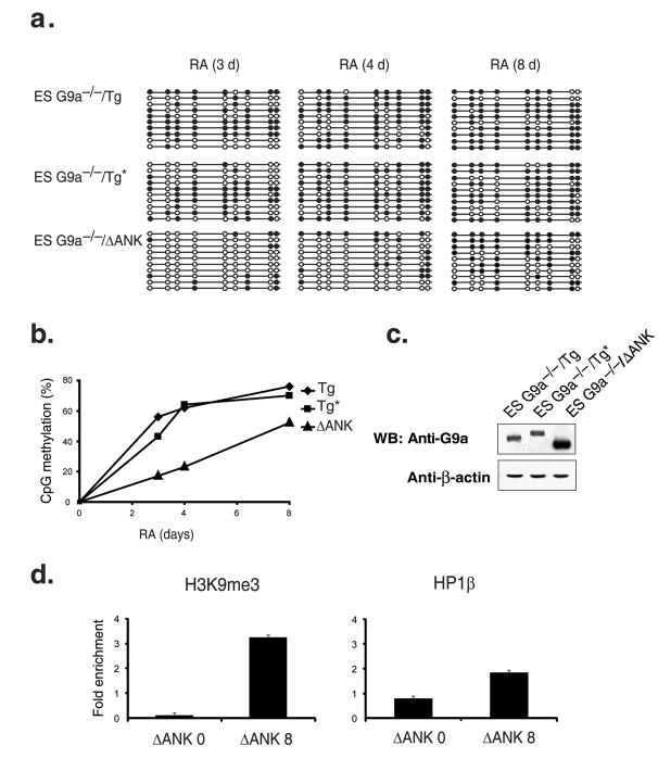
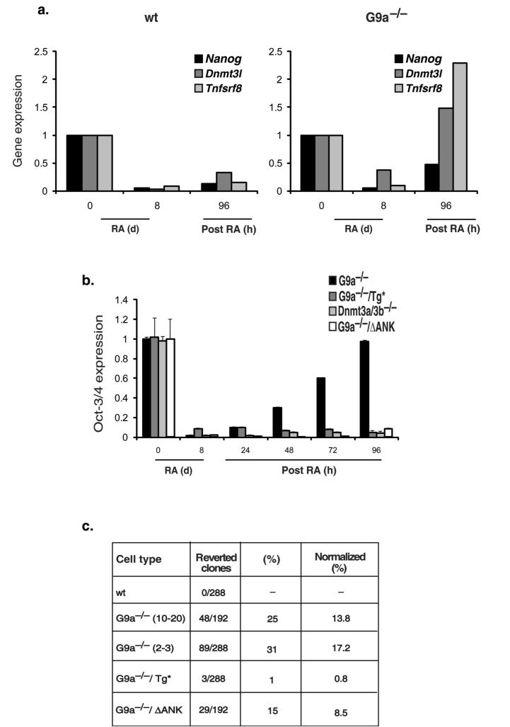
The pluripotency-determining gene Oct3/4 (also called Pou5f1) undergoes postimplantation silencing in a process mediated by the histone methyltransferase G9a. Microarray analysis now shows that this enzyme may operate as a master regulator that inactivates numerous early-embryonic genes by bringing about heterochromatinization of methylated histone H3K9 and de novo DNA methylation. Genetic studies in differentiating embryonic stem cells demonstrate that a point mutation in the G9a SET domain prevents heterochromatinization but still allows de novo methylation, whereas biochemical and functional studies indicate that G9a itself is capable of bringing about de novo methylation through its ankyrin domain, by recruiting Dnmt3a and Dnmt3b independently of its histone methyltransferase activity. These modifications seem to be programmed for carrying out two separate biological functions: histone methylation blocks target-gene reactivation in the absence of transcriptional repressors, whereas DNA methylation prevents reprogramming to the undifferentiated state.


Figures

Figure 1
Figure 1. Genome-wide G9a-dependent de novo methylation
(a) DNA from 8 days RA-treated wt and G9a-/- ES cells and somatic tissues (spleen and kidney) was treated with Na bisulfite (Qiagen bisulfite kit, Cat. No. 59104), amplified using specific gene promoter primers, cloned and subjected to sequence analysis. It should be noted that Nanog de novo methylation was detected at a site different from that measured in previous studies. (b) ChIP analysis of the genes promoters using antibodies specific for H3K9me3 and HP1β on 0 and 8 day RA-treated wt and G9a-/- ES cells.
Figure 2
Figure 2. Heterochromatinization is prevented by point mutation in the G9a SET domain
(a) Chromatin immunoprecipitation (ChIP) analysis of the Oct-3/4 promoter in wild type (wt), G9a-/- and G9a-/-/Tg* (mutated in the SET domain, F1205Y) embryonic stem (ES) cells (RA-differentiated, 8 day) using antibodies specific for H3K9me3, HP1γ and H3K9,14ac. Quantitative real time PCR (Q-PCR) was used. The degree of enrichment was calculated as Bound (B)/Input (I) and normalized to β-actin or β-globin. (b) DNA from 8 day RA-treated ES cells was treated with Na bisulfite, amplified using specific promoter primers (Supplementary Table 2), cloned and subjected to sequence analysis. Nine and three CpG sites, respectively, were examined. (c) ChIP analysis of the Oct-3/4 promoter using antibodies specific for Dnmt3a and Dnmt3b on 6 day differentiated ES cells. Enrichment values were normalized to pericentric satellite DNA in each sample (±SD).
Figure 3
Figure 3. G9a recruits Dnmt3a and Dnmt3b
(a) 293 cells transiently transfected with Dnmt3a or Dnmt3b and G9a-Flag tagged, or with HA-G9a and Gal4-Dnmt3a (as indicated). Cell extracts were immuno-precipitated (IP) with IgG, specific antibodies against Flag or Gal4, and Western blot (WB) analysis performed with anti-Flag, anti-Dnmt3a, anti-Dnmt3b, anti-HA or anti-G9a, as indicated. (b) Direct interactions between G9a and Dnmts. A GST-fused G9a protein was incubated either with His-Dnmt3a or with His-Dnmt3b proteins. WB analysis was performed with anti-His. (c) Endogenous co-immunoprecipitations of G9a and Dnmts. Nuclear extracts prepared from differentiated ES cells were immunoprecipitated with anti-IgG, anti-G9a, or anti-Dnmt3b. WB analysis was performed with anti-Dnmt3b or anti-G9a.
Figure 4
Figure 4. Mapping the G9a- and Dnmts-interacting domains
(a) Schematic representation of the plasmids used for the IP assay. 293 cells transiently transfected with various mutants of G9a EGFP-tagged plasmids. Extracts were immuno-precipitated with anti-EGFP and WB with antibodies specific for EGFP, Dnmt3a and Dnmt3b. (b) Schematic representation of GST-fusion constructs harboring full length Dnmt3L, different regions of Dnmt3a and 3b. The methyltransferase domain is labeled as MTD. In vitro translated (IVT) G9a was incubated with the indicated GST fusions of Dnmt3a, 3b and 3L. The presence of G9a was visualized by WB analysis using anti-G9a. Expression of GST-Dnmts was assayed by SDS-PAGE gels stained with Coomasie-blue.
Figure 5
Figure 5. ANK-dependent Oct-3/4 methylation
(a) Na bisulfite analysis of DNA from G9a-/- ES cells harboring the indicated G9a transgene constructs. (b) The data in (a) is redrawn in graphic form. (c) Cell extracts from transfected ES cells were Western blotted with antibodies specific for G9a and β-actin. (d) ChIP analysis of the Oct-3/4 promoter using anti-H3K9me3 or anti-HP1β on chromatin from G9a-/-/ΔANK ES cells at 0 time and 8 days of RA-induced differentiation.
Figure 6
Figure 6. Reactivation and reprogramming of Oct-3/4, Nanog, Dnmt3L and Tnfsrf8 genes are affected by their epigenetic state
(a) wt and G9a-/- ES cells were treated with RA during 8 days, after which RA was removed and cells were isolated after 96 hours. Graph shows the level of expression for the indicated genes using Q-PCR analysis with Ubiquitin C (UBC) as the normalization control. (b) ES cells were treated with RA for up to 8 days, after which RA was removed and cells were isolated at various time points. Graph shows the level of Oct-3/4 expression (± SD) using Q-PCR analysis with UBC as the normalization control. (c) Number and percentage of reverted single-cell clones established from wild type (wt) and mutant ES cells that were initially differentiated with RA for 8 days and re-cultured in 96-well plates for 2 weeks in the presence of LIF under conditions that were calibrated for each line to yield approximately one colony per well of 1 day RA-treated cells (10-20 cells per well). Each result represents the sum of 2-3 independent experiments. Normalization takes into consideration the clonability of each cell line, as determined by the number of colonies found after plating 1 day RA-treated cells.

References

    1. Surani MA, Hayashi K, Hajkova P. Genetic and epigenetic regulators of pluripotency. Cell. 2007;128:747–762. - PubMed
    1. Reik W. Stability and flexibility of epigenetic gene regulation in mammalian development. Nature. 2007;447:425–432. - PubMed
    1. Niwa H. Open conformation chromatin and pluripotency. Genes Dev. 2007;21:2671–2676. - PubMed
    1. Niwa H, Miyazaki J, Smith AG. Quantitative expression of Oct-3/4 defines differentiation, dedifferentiation or self-renewal of ES cells. Nature Genet. 2000;24:372–376. - PubMed
    1. Chambers I, et al. Functional expression cloning of nanog, a pluripotency sustaining factor in embryonic stem cells. Cell. 2003;113:643–655. - PubMed

Publication types

MeSH terms