. 2008 Dec;4(12):758-65.
doi: 10.1038/nchembio.121. Epub 2008 Oct 26.

Reconstitution of ThiC in thiamine pyrimidine biosynthesis expands the radical SAM superfamily

Affiliations

Reconstitution of ThiC in thiamine pyrimidine biosynthesis expands the radical SAM superfamily

Abhishek Chatterjee et al. Nat Chem Biol. 2008 Dec.

Abstract

4-Amino-5-hydroxymethyl-2-methylpyrimidine phosphate (HMP-P) synthase catalyzes a complex rearrangement of 5-aminoimidazole ribonucleotide (AIR) to form HMP-P, the pyrimidine moiety of thiamine phosphate. We determined the three-dimensional structures of HMP-P synthase and its complexes with the product HMP-P and a substrate analog imidazole ribotide. The structure of HMP-P synthase reveals a homodimer in which each protomer comprises three domains: an N-terminal domain with a novel fold, a central (betaalpha)(8) barrel and a disordered C-terminal domain that contains a conserved CX(2)CX(4)C motif, which is suggestive of a [4Fe-4S] cluster. Biochemical studies have confirmed that HMP-P synthase is iron sulfur cluster-dependent, that it is a new member of the radical SAM superfamily and that HMP-P and 5'-deoxyadenosine are products of the reaction. Mössbauer and EPR spectroscopy confirm the presence of one [4Fe-4S] cluster. Structural comparisons reveal that HMP-P synthase is homologous to a group of adenosylcobalamin radical enzymes. This similarity supports an evolutionary relationship between these two superfamilies.


Figures
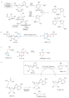
Figure 1
Figure 1
The biosynthesis of thiamin pyrophosphate (a) Overall bacterial pathway. Aminoimidazole ribonucleotide 12 is converted to 4-amino-5-hydroxymethyl-2-methylpyrimidine phosphate 15 by HMP-P synthase (ThiC), which is phosphorylated by ThiD to give HMP pyrophosphate 3. The thiazole moiety 2 is biosynthesized from 1-deoxy-D-xyulose 5-phosphate 6, cysteine 8 and dehydroglycine 35. The dehydroglycine is generated from glycine (ThiO) in B. subtilis and from tyrosine (ThiH) in E. coli. The pyrimidine and thiazole are coupled by ThiE to give thiamin phosphate 4 and ThiL catalyzes the final phosphorylation. (b) Conversion of AIR 12 to the thiamin pyrimidine in bacteria and plants. The color coding indicates the source of non-hydrogen atoms in HMP-P as demonstrated by labeling studies. (c) Biosynthesis of thiamin pyrimidine in fungi. In fungi the pyrimidine moiety is derived from histidine 13 and pyridoxal 5′-phosphate 14 using a single enzyme, THI5p. The color coding indicates the source of non-hydrogen atoms. (d) The HMP-P synthase reactions. When Fe-S cluster loaded HMP-P synthase is reduced with dithionite, it reduces SAM 16 to generate methionine 28 and the 5′-deoxyadenosine (5-dAdo) radical 17, which is required by HMP-P synthase to convert AIR 12 to HMP-P 15.
Figure 2
Figure 2
HMP-P synthase activity. (a) UV-Visible absorption spectrum of isolated, Fe-S cluster reconstituted, HMP-P synthase (25 μM; black trace) and its change upon reduction with 200 μM dithionite (gray trace). (b) In vitro reconstitution of HMP-P biosynthesis, analyzed by HPLC after converting HMP-P 15 to the fluorescent thiochrome phosphate 19. Enhanced product yield is observed when HMP-P synthase is subjected to chemical Fe-S cluster reconstitution. Blue and black traces represent reactions utilizing Fe-S cluster reconstituted and untreated HMP-P synthase lysate respectively; red trace represents a reaction lacking AIR 12. The thiochrome pyrophosphate 20 peak arises from thiamin pyrophosphate in lysate and stays constant. (c) Activity of Fe-S cluster reconstituted, purified HMP-P synthase. Sample 1 is reaction mixture containing HMP-P synthase, AIR, SAM, dithionite. Sample 2 is reaction 1 incubated aerobically. Samples 3, 4 and 5 are reaction 1 without SAM, AIR and dithionite respectively. (d) HPLC analysis of the HMP-P synthase reactions. Blue trace represents full reaction [HMP-P synthase, AIR, SAM and dithionite]; green trace, red trace and black trace represent the full reaction without AIR, SAM and dithionite respectively. Reaction products HMP-P 15 (rt = 2.5 min) and 5-dAdo 18 (rt = 18.45 min) were identified by comigration with authentic standards and subsequent spectroscopic characterization. Three sections of the chromatogram (1.5–2.7 min, 5–6 min, 18.3–18.6 min) are presented to clearly demonstrate the changes.
Figure 3
Figure 3
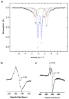
EPR and Mossbauer spectroscopy of HMP-P synthase. (a) 4.2-K/53-mT Mössbauer spectrum of a sample of HMP-P synthase overexpressed in bacteria grown on 57Fe-supplemented media and further reconstituted with 57Fe, sulfide, and DTT (hashed marks). The solid lines are simulations with three quadrupole doublets using the parameters quoted in the text, representing [4Fe-4S]2+ clusters (blue), and two distinct high-spin FeII species (red and green). (b) EPR spectrum of an as-isolated sample of HMP-P synthase that was coexpressed with Isc cluster. Presence of an organic radical is noted with parameters mentioned in the text. (c) EPR spectrum of reconstituted HMP-P synthase. The sample (500 μM) was reduced with 10 mM dithionite before anaerobically loading into an EPR tube and freezing. The spectrum was obtained under the following conditions: microwave power, 101 μW; receiver gain, 3; modulation amplitude, 10 G; temperature, 13 K; microwave frequency, 9.5 GHz.
Figure 4
Figure 4
Structure of HMP-P synthase. (a) The HMP-P synthase homodimer. The protomer consists of three domains. The N-terminal domains are colored in shades of blue, the (βα)8 core domains are colored in shades of green and the C-terminal domains are colored in shades of red. HMP-P is shown as ball-and-stick. The final 66 amino acids are disordered; however, the final ordered residues, which immediately precede a conserved CX2CX4C motif, extend into the active site of the adjacent protomer. The C-terminal tail is anchored to the adjacent protomer by a three helix bundle motif located at the beginning of the C-terminal domain. (b) Stereoview of the HMP-P synthase active site with modeled SAM and the [4Fe-4S] cluster. The atoms are color coded by atom type (green = C, blue = N, red = O, yellow = S and orange = Fe). The substrate analog IMR 22 from the crystal structure is shown. Residues Cys561, Cys564 and Cys 569, SAM and the [4Fe-4S] cluster were modeled using biotin synthase as a guide. Hydrogen bonds are indicated by dotted lines. (c) Superposition of the (βα)8 domains from HMP-P synthase and biotin synthase (PDB ID 1r3o). HMP-P synthase is shown in blue and biotin synthase is shown in silver. The [4Fe-4S] cluster and SAM from biotin synthase are shown in ball-and-stick.
Figure 5
Figure 5
Cartoons depicting the domain assemblies of cobalamin-dependent enzymes with HMP-P synthase-like protomers and dimer interfaces. Each chain within a molecule is color coded differently. (a) Glutamate mutase from C. sticklandii. GM contains two identical catalytic subunits and two identical AdoCbl binding subunits, which cap the catalytic domains, forming an (AB)2 heterotetramer. (b) Lysine 5,6-amino mutase. 5,6-LAM forms an (AB)2 heterotetramer with an assembly similar to that of GM. (c) Methylmalonyl coenzyme A mutase from P. shermanii. The catalytic and AdoCbl binding domains are fused (A). The blue subunit (B) does not contain a catalytic site. (d) Carbon monoxide dehydrogenase corrinoid/iron-sulfur protein. The blue subunit contains a (βα)8 domain (B), with no catalytic site, fused to an MeCbl binding domain, which caps the active site of the catalytic domain (A). (e) HMP-P synthase from C. crescentus. HMP synthase is a homodimer. The (βα)8 core domain is fused to the predicted [4Fe-4S] binding domain. This domain is disordered in the crystal structure but the final ordered residues extend into the active site of the adjacent protomer and are preceded by a three helix bundle that anchors the C-terminus to the adjacent protomer.

References

    1. Begley TP, et al. Thiamin biosynthesis in prokaryotes. Arch Microbiol. 1999;171:293–300. - PubMed
    1. Settembre E, Begley TP, Ealick SE. Structural biology of enzymes of the thiamin biosynthesis pathway. Curr Opin Struct Biol. 2003;13:739–747. - PubMed
    1. Chatterjee A, Jurgenson CT, Schroeder FC, Ealick SE, Begley TP. Thiamin Biosynthesis in Eukaryotes: Characterization of the Enzyme-Bound Product of Thiazole Synthase from Saccharomyces cerevisiae and Its Implications in Thiazole Biosynthesis. J Am Chem Soc. 2006;128:7158–7159. - PMC - PubMed
    1. Chatterjee A, Jurgenson CT, Schroeder FC, Ealick SE, Begley TP. Biosynthesis of Thiamin Thiazole in Eukaryotes: Conversion of NAD to an Advanced Intermediate. J Am Chem Soc. 2007;129:2914–2922. - PMC - PubMed
    1. Jurgenson CT, Chatterjee A, Begley TP, Ealick SE. Structural Insights into the Function of the Thiamin Biosynthetic Enzyme Thi4 from Saccharomyces cerevisiae. Biochemistry. 2006;45:11061–11070. - PubMed

Publication types

MeSH terms

Associated data