. 2008;3(10):e3517.
doi: 10.1371/journal.pone.0003517. Epub 2008 Oct 27.

Wnt5a regulates ventral midbrain morphogenesis and the development of A9-A10 dopaminergic cells in vivo

Affiliations

Wnt5a regulates ventral midbrain morphogenesis and the development of A9-A10 dopaminergic cells in vivo

Emma R Andersson et al. PLoS One. 2008.

Abstract

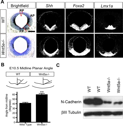
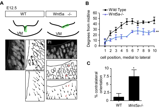
Wnt5a is a morphogen that activates the Wnt/planar cell polarity (PCP) pathway and serves multiple functions during development. PCP signaling controls the orientation of cells within an epithelial plane as well as convergent extension (CE) movements. Wnt5a was previously reported to promote differentiation of A9-10 dopaminergic (DA) precursors in vitro. However, the signaling mechanism in DA cells and the function of Wnt5a during midbrain development in vivo remains unclear. We hereby report that Wnt5a activated the GTPase Rac1 in DA cells and that Rac1 inhibitors blocked the Wnt5a-induced DA neuron differentiation of ventral midbrain (VM) precursor cultures, linking Wnt5a-induced differentiation with a known effector of Wnt/PCP signaling. In vivo, Wnt5a was expressed throughout the VM at embryonic day (E)9.5, and was restricted to the VM floor and basal plate by E11.5-E13.5. Analysis of Wnt5a-/- mice revealed a transient increase in progenitor proliferation at E11.5, and a precociously induced NR4A2+ (Nurr1) precursor pool at E12.5. The excess NR4A2+ precursors remained undifferentiated until E14.5, when a transient 25% increase in DA neurons was detected. Wnt5a-/- mice also displayed a defect in (mid)brain morphogenesis, including an impairment in midbrain elongation and a rounded ventricular cavity. Interestingly, these alterations affected mostly cells in the DA lineage. The ventral Sonic hedgehog-expressing domain was broadened and flattened, a typical CE phenotype, and the domains occupied by Ngn2+ DA progenitors, NR4A2+ DA precursors and TH+ DA neurons were rostrocaudally reduced and laterally expanded. In summary, we hereby describe a Wnt5a regulation of Wnt/PCP signaling in the DA lineage and provide evidence for multiple functions of Wnt5a in the VM in vivo, including the regulation of VM morphogenesis, DA progenitor cell division, and differentiation of NR4A2+ DA precursors.


Conflict of interest statement

Competing Interests: The authors have declared that no competing interests exist.

Figures

Figure 1
Figure 1. Temporal and spatial expression of Wnt5a in the mouse.
(A) In situ hybridization for Wnt5a on coronal midbrain sections of E9.5–E13.5 CD1 mice shows a dynamic regulation of Wnt5a expression domains throughout development in both rostrocaudal distribution and developmental stages. Schemes with sagittal sections of the brain and dashed lines show the levels at which rostral (r) or caudal (c) expression analysis was performed. The expression of Wnt5a occupies the entire neuroepithelium of the ventral midbrain at E9.5 and 10.5, and becomes progressively restricted to the floor plate and basal plate ventricular zone from E11.5–E13.5, extending to the marginal zone at caudal levels (Scale bars at E9.5 = 100 µm, at E10.5 and E11.5 = 200 µm, at E12.5 and E13.5 = 175 µm). (B) At E12.5, the expression of Wnt5a comprises the FP extending from the ventricular zone through the intermediate zone and into the marginal zone, overlapping with NR4A2+ and TH+ cells. This extension into the marginal zone is most pronounced in the caudal midbrain. (C) The expression of Wnt5a is down-regulated by E18.5, where it still overlaps with TH+ cells, but 8 weeks after birth, at P56, this overlap is lost as seen in sagittal and coronal sections. (Scale bars in brightfield E18.5, coronal P56 and darkfield sagital p56 = 500 µm, scale bar in brightfield sagital P56 = 2.5 mm). Abbreviations: r = rostral, c = caudal, F = forebrain, D = diencephalon, M = midbrain, H = hindbrain.
Figure 2
Figure 2. Rac1 is activated by Wnt5a and is required for Wnt5a-induced dopaminergic neuron differentiation.
(A) Stimulation of SN4741 cells, a midbrain dopaminergic neuron cell line, with 200 ng/ml Wnt5a for 2 hrs activates Rac1, but not RhoA or Cdc42. (B) As previously reported, Wnt5a upregulates the number of TH+ dopaminergic neurons in E11.5 primary ventral midbrain cultures grown for three days in vitro. This differentiation is completely inhibited in the presence of 10 µM NSC23766, a Rac1 inhibitor (ANOVA for Wnt5a vs Wnt5a&NSC23766, P<0.05, N = 3 experiments, each normalized to the control conditions) (C) TH+ cells in all conditions have normal morphology, with increased numbers in the Wnt5a-treated, but not Rac-inhibited, wells.
Figure 3
Figure 3. The number of dopaminergic neurons is normal at most stages but transiently increases at E14.5 in Wnt5a−/− mice.
(A) At E11.5 and E12.5 no differences in the number of TH+ cells could be detected. At E12.5 and E14.5 the region occupied by TH+ cells in the Wnt5a−/−midbrain appears larger, extending both laterally and dorsally. (B) At E17.5 the distribution of cells is broader dorsoventrally in rostral and caudal sections in the Wnt5a−/− VM. Importantly, at rostral levels the ventral tegmental area was more lateral (leaving a TH-poor midline domain) and the substantia nigra more medial, making it difficult to differentiate between them. (C) Enlarged image of TH+ cells at E12.5 shows normal DA neuron morphology in the Wnt5a−/− mice. (D) Quantification of the number of TH+ cells in E11.5, E12.5, E14.5, and E17.5 mice shows a transient 25% increase at E14.5 in Wnt5a−/− embryos, which was no longer seen at E17.5. (At E14.5, unpaired t-test, p = 0.0492, N = 4).
Figure 4
Figure 4. Increased proliferation and accumulation of progenitor cells in Wnt5a−/− mice.
(A). Representative coronal sections at the level of the midbrain of 2 hr BrdU-pulsed WT and Wnt5a−/− embryos at E11.5 immunostained for BrdU or Ki67, markers of cells in mitosis. The black squares in (A) depict the area within the alar plate (AP) and floor plate (FP) used for the quantification of BrdU+ cells in (B) (Squares in (A) are not drawn to scale). Note the accumulation of BrdU+ cells in the Wnt5a−/− VM in the adjacent magnified box. Increased Ki67 staining and mitotic figures (arrowheads) in the ventricular zone. (B) The density of BrdU+ proliferating cells was significantly increased within the FP, but no significant change was found within the AP of Wnt5a−/− embryos compared to their wt littermates (C) The number of Ki67+ cells was significantly increased at E12.5 (paired t-test, p = 0.003, N = 3). (D) Mitotic nuclei (arrowheads in (A)) were counted in the midline domain to asses the number of cells in M-phase, and this was also found to be significantly increased in the Wnt5a−/− mutant (paired t-test, p = 0.037, N = 3).
Figure 5
Figure 5. The ventral midbrain progenitor domains are expanded in Wnt5a−/− embryos.
(A) Detection of Th, Ngn1, and Ngn2 on representative serial coronal sections at the level of the midbrain of WT and Wnt5a−/− embryos at E11.5. Red brackets delimit the ventral Ngn1 and Ngn2 domain in the WT embryo, respectively. (Scale bar in A: 500 µm.). (B) Increase in NR4A2+ cells in the VM of Wnt5a−/− mice, which occupy a broader region laterally and dorsoventrally. (C) A 47% increase in the number of NR4A2+ cells was detected in the Wnt5a−/− mice at E12.5 (unpaired t-test, p = 0.044, WT N = 3, Wnt5a−/− N = 4). (D) Analysis of the proportion of NR4A2+ cells differentiating into TH+ DA neurons revealed that the differentiation of NR4A2+ precursors into TH+ cells was impaired in the Wnt5a−/− mice at E12.5 (paired t-test, p = 0.0098, N = 3).
Figure 6
Figure 6. Lateral expansion of Sonic hedgehog expression and flattening of midbrain ventricle, associated with loss of N-cadherin in Wnt5a−/− embryos.
(A) Shh, Foxa2 and Lmx1a are laterally expanded by E11.5 in Wnt5a−/− mice. The invagination at the medial hinge-point is markedly reduced/flattened in the mutants. Red bars delimit the alar plate (AP), basal plate (BP), floor plate (FP) and roof plate (RP). Scale bar 250 µm. (B) The flattened VM invagination was quantified by measuring the angle formed between the midline and the ventricular wall at several levels throughout the VM of E10.5 mice, when this morphological change first became obvious. A significant difference was found in the Wnt5a−/− mice forming a U-shaped ventricle, with an angle of 59.4°±2.8° from the midline as compared to 49.1°±4.3° in WT mice. (C) Western blot of E9.5 whole brain of WT and Wnt5a−/− mice revealed a marked reduction of N-cadherin in Wnt5a−/− mice.
Figure 7
Figure 7. Apical-basal polarity/cell orientation are affected in Wnt5a−/− mice.
(A) At E12.5, propidium iodide staining on coronal sections through the VM revealed that cell nuclei in the neuroepithelium were rounded and their orientation was more variable with some cells pointing contralaterally (red asterisks/arrows). (B) The orientation of each cell nucleus was plotted versus its distance from the ventral midline. The angle between the nucleus and the midline was measured from cell 1 (the most medial) to cell 10 (the most lateral). Cell nuclei in Wnt5a−/− mice are oriented more ventrally compared to the more lateral orientation of cells in WT mice (two way-ANOVA for genotype and level, p = 0.0029, N = 3). (C) The frequency of cell nuclei oriented towards the contralateral ventral side (red arrows in A) was significantly increased in Wnt5a−/− mice (paired t-test, p = 0.0198, N = 3, 10 nuclei at 3 levels/animal).
Figure 8
Figure 8. The distribution of TH+ cells in the rostrocaudal and mediolateral axis is altered in Wnt5a−/− mice.
(A) At E12.5, there is a redistribution of TH+ cells in the VM, both in the rostrocaudal and mediolateral axis. (B) Three representative levels throughout the VM were measured for lateral spread of TH+ cells: rostral, medial and caudal. ImageJ was used to measure the distance between the two TH+ cells furthest apart in (D). (C) TH+ cells were quantified at regular intervals from rostral to caudal midbrain in (E–F). (D) TH+ cells are distributed much more broadly in anterior midbrain of E12.5 Wnt5a−/− mice compared to WT (ANOVA, N = 3, p<0.001). (ANOVA with Bonferroni's post test, Rostral WT vs Rostral Wnt5a−/− p<0.001). (E–F) Rostrocaudal distribution of TH+ cells. (E) At E11.5 no difference could be seen between the distribution of TH+ cells in the anteroposterior axis of WT or Wnt5a−/− mice. However, at E12.5 (F) and E14.5 (G) an altered distribution with a decrease in the number of TH+ cells in anterior levels and an increase in medial levels was seen (Two way ANOVA, for level and genotype, P = 0.0016 at E12.5, P<0.0001 at E14.5, N = 4). (H) The lateral expansion of TH+ cells at E12.5 was confirmed by in situ hybridization for Th, Pitx3 and DAT, two other markers of maturing dopaminergic neurons.

References

    1. Clevers H. Wnt/beta-catenin signaling in development and disease. Cell. 2006;127:469–480. - PubMed
    1. Seifert JR, Mlodzik M. Frizzled/PCP signalling: a conserved mechanism regulating cell polarity and directed motility. Nat Rev Genet. 2007;8:126–138. - PubMed
    1. Mikels AJ, Nusse R. Purified Wnt5a protein activates or inhibits beta-catenin-TCF signaling depending on receptor context. PLoS Biol. 2006;4:e115. - PMC - PubMed
    1. Qian D, Jones C, Rzadzinska A, Mark S, Zhang X, et al. Wnt5a functions in planar cell polarity regulation in mice. Dev Biol. 2007;306:121–133. - PMC - PubMed
    1. Witze ES, Litman ES, Argast GM, Moon RT, Ahn NG. Wnt5a control of cell polarity and directional movement by polarized redistribution of adhesion receptors. Science. 2008;320:365–369. - PMC - PubMed

Publication types

MeSH terms