Osteoclasts control osteoblast chemotaxis via PDGF-BB/PDGF receptor beta signaling
- PMID: 18953417
- PMCID: PMC2569415
- DOI: 10.1371/journal.pone.0003537
Osteoclasts control osteoblast chemotaxis via PDGF-BB/PDGF receptor beta signaling
Abstract
Background: Bone remodeling relies on the tightly regulated interplay between bone forming osteoblasts and bone digesting osteoclasts. Several studies have now described the molecular mechanisms by which osteoblasts control osteoclastogenesis and bone degradation. It is currently unclear whether osteoclasts can influence bone rebuilding.
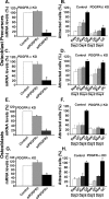
Methodology/principal findings: Using in vitro cell systems, we show here that mature osteoclasts, but not their precursors, secrete chemotactic factors recognized by both mature osteoblasts and their precursors. Several growth factors whose expression is upregulated during osteoclastogenesis were identified by DNA microarrays as candidates mediating osteoblast chemotaxis. Our subsequent functional analyses demonstrate that mature osteoclasts, whose platelet-derived growth factor bb (PDGF-bb) expression is reduced by siRNAs, exhibit a reduced capability of attracting osteoblasts. Conversely, osteoblasts whose platelet-derived growth factor receptor beta (PDGFR-beta) expression is reduced by siRNAs exhibit a lower capability of responding to chemotactic factors secreted by osteoclasts.
Conclusions/significance: We conclude that, in vitro mature osteoclasts control osteoblast chemotaxis via PDGF-bb/PDGFR-beta signaling. This may provide one key mechanism by which osteoclasts control bone formation in vivo.
Conflict of interest statement
Figures


Similar articles
-
Platelet-derived growth factor BB enhances osteoclast formation and osteoclast precursor cell chemotaxis.J Bone Miner Metab. 2017 Jul;35(4):355-365. doi: 10.1007/s00774-016-0773-8. Epub 2016 Sep 14. J Bone Miner Metab. 2017. PMID: 27628046
-
Platelet-derived growth factor BB secreted from osteoclasts acts as an osteoblastogenesis inhibitory factor.J Bone Miner Res. 2002 Feb;17(2):257-65. doi: 10.1359/jbmr.2002.17.2.257. J Bone Miner Res. 2002. PMID: 11811556
-
Non-resorbing osteoclasts induce migration and osteogenic differentiation of mesenchymal stem cells.J Cell Biochem. 2010 Feb 1;109(2):347-55. doi: 10.1002/jcb.22406. J Cell Biochem. 2010. PMID: 19950208
-
Osteoclasts Provide Coupling Signals to Osteoblast Lineage Cells Through Multiple Mechanisms.Annu Rev Physiol. 2020 Feb 10;82:507-529. doi: 10.1146/annurev-physiol-021119-034425. Epub 2019 Sep 25. Annu Rev Physiol. 2020. PMID: 31553686 Review.
-
Osteoblast migration in vertebrate bone.Biol Rev Camb Philos Soc. 2018 Feb;93(1):350-363. doi: 10.1111/brv.12345. Epub 2017 Jun 19. Biol Rev Camb Philos Soc. 2018. PMID: 28631442 Free PMC article. Review.
Cited by
-
Wnt Signaling Inhibits Osteoclast Differentiation by Activating Canonical and Noncanonical cAMP/PKA Pathways.J Bone Miner Res. 2016 Jan;31(1):65-75. doi: 10.1002/jbmr.2599. Epub 2015 Aug 19. J Bone Miner Res. 2016. PMID: 26189772 Free PMC article.
-
Nervous System-Driven Osseointegration.Int J Mol Sci. 2022 Aug 10;23(16):8893. doi: 10.3390/ijms23168893. Int J Mol Sci. 2022. PMID: 36012155 Free PMC article. Review.
-
Breast cancer metastasis to the bone: mechanisms of bone loss.Breast Cancer Res. 2010;12(6):215. doi: 10.1186/bcr2781. Epub 2010 Dec 16. Breast Cancer Res. 2010. PMID: 21176175 Free PMC article. Review.
-
New Bone Formation in Tuberculous-Infected Vertebral Body Defect after Administration of Bone Marrow Stromal Cells in Rabbit Model.Asian Spine J. 2016 Feb;10(1):1-5. doi: 10.4184/asj.2016.10.1.1. Epub 2016 Feb 16. Asian Spine J. 2016. PMID: 26949451 Free PMC article.
-
Citric Acid: A Nexus Between Cellular Mechanisms and Biomaterial Innovations.Adv Mater. 2024 Aug;36(32):e2402871. doi: 10.1002/adma.202402871. Epub 2024 Jun 11. Adv Mater. 2024. PMID: 38801111 Free PMC article. Review.
References
-
- Teitelbaum SL. Bone resorption by osteoclasts. Science. 2000;289:1504–1508. - PubMed
-
- Aguila HL, Rowe DW. Skeletal development, bone remodeling, and hematopoiesis. Immunol Rev. 2005;208:7–18. - PubMed
-
- Boyle WJ, Simonet WS, Lacey DL. Osteoclast differentiation and activation. Nature. 2003;423:337–342. - PubMed
-
- Kawamata A, Izu Y, Yokoyama H, Amagasa T, Wagner EF, et al. JunD suppresses bone formation and contributes to low bone mass induced by estrogen depletion. J Cell Biochem. 2008;103:1037–1045. - PubMed
Publication types
MeSH terms
Substances
LinkOut - more resources
Full Text Sources
Other Literature Sources
Molecular Biology Databases
Miscellaneous

