. 2008 Nov;10(11):1204-12.
doi: 10.1593/neo.08642.

The neurofibromatosis 2 tumor suppressor gene product, merlin, regulates human meningioma cell growth by signaling through YAP

Affiliations
Free PMC article

The neurofibromatosis 2 tumor suppressor gene product, merlin, regulates human meningioma cell growth by signaling through YAP

Katherine Striedinger et al. Neoplasia. 2008 Nov.
Free PMC article

Abstract

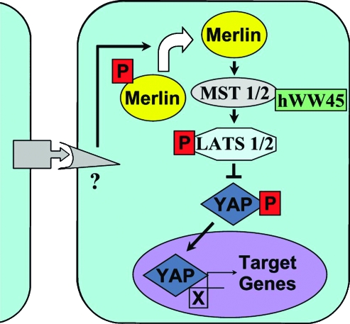
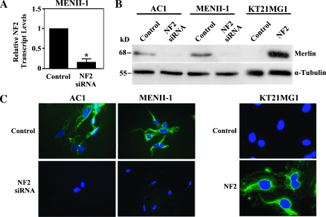
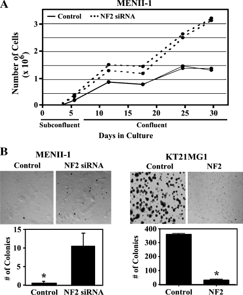
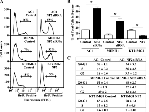
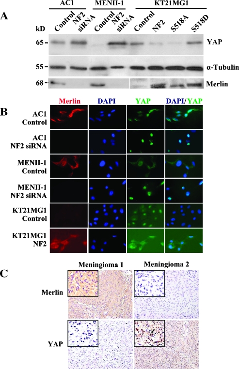
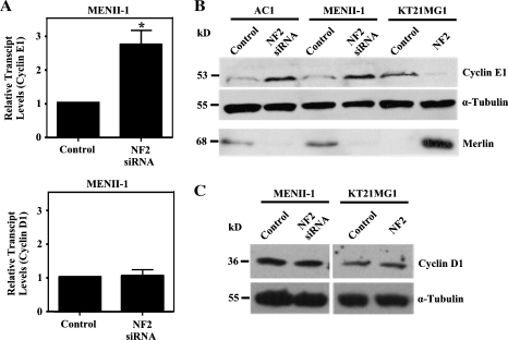
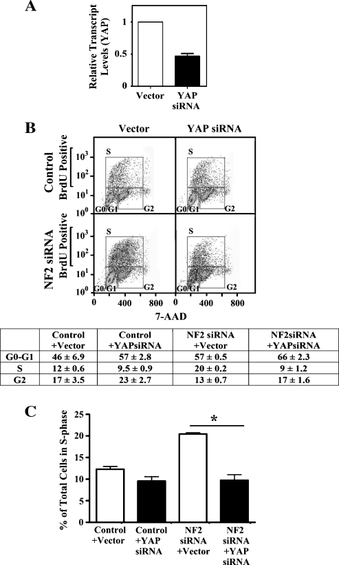
Neurofibromatosis type 2 (NF2) is an autosomal dominant disorder characterized by the occurrence of schwannomas and meningiomas. Several studies have examined the ability of the NF2 gene product, merlin, to function as a tumor suppressor in diverse cell types; however, little is known about merlin growth regulation in meningiomas. In Drosophila, merlin controls cell proliferation and apoptosis by signaling through the Hippo pathway to inhibit the function of the transcriptional coactivator Yorkie. The Hippo pathway is conserved in mammals. On the basis of these observations, we developed human meningioma cell lines matched for merlin expression to evaluate merlin growth regulation and investigate the relationship between NF2 status and Yes-associated protein (YAP), the mammalian homolog of Yorkie. NF2 loss in meningioma cells was associated with loss of contact-dependent growth inhibition, enhanced anchorage-independent growth and increased cell proliferation due to increased S-phase entry. In addition, merlin loss in both meningioma cell lines and primary tumors resulted in increased YAP expression and nuclear localization. Finally, siRNA-mediated reduction of YAP in NF2-deficient meningioma cells rescued the effects of merlin loss on cell proliferation and S-phase entry. Collectively, these results represent the first demonstration that merlin regulates cell growth in human cancer cells by suppressing YAP.


Figures

Figure 1
Figure 1
Schematic of the mammalian Hippo signaling pathway. The Hippo pathway is an evolutionary conserved cellular pathway that coordinately regulates cell proliferation and apoptosis. Merlin has been proposed to interact with unknown membrane proteins and transduce a signal that stimulates the phosphorylation of LATS1/2 by the serine/threonine kinases MST1/2 that interact with hWW45. LATS1/2 inhibits the transcriptional coactivator YAP resulting in suppressed expression of downstream target genes such as cyclins that are involved in cell growth and proliferation.
Figure 2
Figure 2
In vitro model system of merlin expression in human meningeal cells. Endogenous merlin was silenced in AC1 and MENII-1 cells using NF2 specific siRNA. In parallel, merlin isoform 1 was exogenously expressed in KT21MG1 cells using retroviral mediated gene transfer. (A) NF2 transcript levels were measured by quantitative PCR and showed a 5.6-fold reduction in MENII-1-NF2-siRNA cells compared with MENII-1-Control cells. Asterisk denotes statistical significance (P < .05). (B) Western blot analysis of cell lysates derived from AC1, MENII-1, and KT21MG1 stable cell populations was used to confirm loss or gain of merlin. Whereas merlin expression was observed in AC1-Control and MENII-1-Control cells, NF2 siRNA abolished expression of merlin in AC1-NF2-siRNA and MENII-1-NF2-siRNA cells. In parallel, KT21MG1-Control cells lacked merlin, whereas KT21MG1-NF2 cells expressed wild type merlin. Levels of α-tubulin were determined in the same samples as a loading control. Immunoblot of one representative experiment of three with similar results is shown. (C) Immunofluorescence using the A19 polyclonal antibody against merlin revealed the presence of cytoplasmic staining in AC1-Control and MENII-1-Control cells and its absence in AC1-NF2-siRNA and MENII-1-NF2-siRNA cells. In contrast, KT21MG1-Control cells had no staining, whereas KT21MG1-NF2 cells had cytoplasmic staining. Merlin immunolabeling is shown in green, and nuclear DAPI counterstaining is shown in blue.
Figure 3
Figure 3
Suppression of merlin causes loss of contact-dependent inhibition of growth and promotes anchorage-independent growth. (A) Growth curves in the presence and absence of merlin expression. Cultures were subconfluent during the first 6 days. MENII-1-NF2-siRNA cells (dotted lines) continued growing after confluent conditions and have less contact-dependent inhibition of growth compared with MENII-1-Control cells (solid lines). Each line corresponds to representative cultures. (B) NF2 suppression promotes anchorage-independent growth. Marked increased in colony formation in soft agar was observed in cells without merlin expression as MENII-1-NF2-siRNA and KT21MG1-Control cells compared with MENII-1-Control and KT21MG1-NF2, respectively. Representative images of the colonies formed (upper panel) and the mean number of colonies per well (lower panel) are shown. Error bars equal ±SE of three independent experiments. Asterisks denote statistical significance (P < .05).
Figure 4
Figure 4
Merlin loss enhances S-phase entry. AC1 and meningioma cell lines (MENII-1 and KT21MG1) were labeled with BrdU and 7-AAD to assess the cell cycle distribution of individual cells by flow cytometry. (A) Representative flow cytometric histograms indicate an increase in the percent of BrdU-positive cells in AC1-NF2-siRNA, MENII-1-NF2-siRNA, and KT21MG1-Control cells compared with AC1-Control, MENII-1-Control, and KT21MG1-NF2 cells, respectively. (B) Bar graphs depict the percentage of cells in the S-phase of the cell cycle (BrdU-positive cells) averaged from three independent experiments. Error bars correspond to ±SE. Asterisks denote statistical significance using unpaired t test (P <.05). (C) Table shows the mean of the percentage of cells ±SE in each phase of the cell cycle from three independent experiments.
Figure 5
Figure 5
Protein levels of YAP are up-regulated and localized to the nucleus in NF2-deficient cells. (A) Total cell lysates were subjected to Western blot using a YAP- or merlin-specific antibody. Increased YAP protein expression was observed when NF2 was suppressed in AC1 and MENII-1 cells compared with controls. Conversely, exogenous expression of merlin decreased YAP in KT21MG1 cells compared with controls. Expression of a nonphosphorylated, active merlin (S518A NF2) was also associated with lower levels of YAP compared with the expression of pseudophosphorylated inactive merlin (S518D NF2). Levels of α-tubulin were determined in the same samples as loading control. Results were reproduced in three independent experiments. (B) YAP was translocated to the nucleus in merlin-deficient cells. Immunofluorescence staining was used to show that YAP was localized to the nucleus in AC1-NF2-siRNA, MENII-1-NF2-siRNA, and KT21MG1-Control cells. In contrast, YAP was primarily cytoplasmic in AC1-Control, MENII-1-Control, and KT21MG1-NF2 cells. Merlin immunolabeling is shown in red; YAP staining is shown in green; and nuclear DAPI counterstaining is shown in blue. (C) In situ immunostaining of merlin and YAP in serial sections of primary human meningioma tumors. We surveyed 37 primary meningiomas by immunohistochemistry. YAP expression was minimal to absent in 95% of merlin-positive meningiomas (a representative tumor is shown here as Meningioma 1). In contrast, YAP was expressed and localized to the nucleus in 92% of merlin-negative meningiomas (a representative tumor is shown here as Meningioma 2). Arrow depicts an example of YAP nuclear localization. Insets show images at higher magnification.
Figure 6
Figure 6
Cyclin E1 and cyclin D1 expression was increased in merlin-deficient cells. (A) Transcript levels of cyclin E1 and cyclin D1 were measured in MENII-1 cells using quantitative PCR. At least a 2.5-fold increase in the transcript levels of cyclin E1 was seen in MENII-1-NF2-siRNA cells compared with MENII-1-Control cells, whereas transcript levels of cyclin D1 were unchanged. Asterisk denotes statistical significance (P < .05). (B) Western blot analysis of cell lysates derived from AC1, MENII-1, and KT21MG1 stable cells was used to show that protein levels of cyclin E1 were increased in the absence of merlin. Levels of α-tubulin were determined in the same samples as a loading control. (C) Western blot analysis of cell lysates derived from MENII-1 and KT21MG1 stable cells was used to show that protein levels of cyclin D1 were unchanged in the absence of merlin. Levels of α-tubulin were determined in the same samples as a loading control.
Figure 7
Figure 7
Down-regulation of YAP decreased proliferation in merlin-deficient cells. MENII-1-Control and MENII-1-NF2-siRNA cells were transiently transfected with YAP-specific siRNA or empty vector. (A) YAP transcript levels were measured in MENII-1-Control cells using quantitative PCR. The YAP-specific siRNA caused a 50% reduction in YAP transcript levels compared with controls in MENII-1 cells. (B, C) BrdU incorporation and 7-AAD staining were measured by flow cytometry. (B) One representative experiment shows the distribution of cells in G0-G1, S, and G2 phases of the cell cycle. The table below shows the mean of the percentage of cells ±SE in each phase of the cell cycle fromthree independent experiments. (C) Bar graphs depict the percentage of cells in the S-phase of the cell cycle (BrdU-positive cells) averaged from three independent experiments. Error bars correspond to ±SE. Suppression of YAP decreased the percentage of cells in S-phase in MENII-1-NF2-siRNA cells to levels similar to MENII-1-Control cells.

References

    1. Ferner RE. Neurofibromatosis 1 and neurofibromatosis 2: a twenty first century perspective. Lancet Neurol. 2007;6:340–351. - PubMed
    1. Ruttledge MH, Sarrazin J, Rangaratnam S, Phelan CM, Twist E, Merel P, Delattre O, Thomas G, Nordenskjold M, Collins VP, et al. Evidence for the complete inactivation of the NF2 gene in the majority of sporadic meningiomas. Nat Genet. 1994;6:180–184. - PubMed
    1. Kalamarides M, Niwa-Kawakita M, Leblois H, Abramowski V, Perricaudet M, Janin A, Thomas G, Gutmann DH, Giovannini M. Nf2 gene inactivation in arachnoidal cells is rate-limiting for meningioma development in the mouse. Genes Dev. 2002;16:1060–1065. - PMC - PubMed
    1. Kalamarides M, Stemmer-Rachamimov AO, Takahashi M, Han ZY, Chareyre F, Niwa-Kawakita M, Black PM, Carroll RS, Giovannini M. Natural history of meningioma development in mice reveals: a synergy of Nf2 and p16 (Ink4a) mutations. Brain Pathol. 2008;18:62–70. - PMC - PubMed
    1. Gautreau A, Louvard D, Arpin M. ERM proteins and NF2 tumor suppressor: the yin and yang of cortical actin organization and cell growth signaling. Curr Opin Cell Biol. 2002;14:104–109. - PubMed

Publication types

MeSH terms