. 2008 Dec;466(12):2981-94.
doi: 10.1007/s11999-008-0556-1. Epub 2008 Oct 25.

Periosteal grafting for congenital pseudarthrosis of the tibia: a preliminary report

Affiliations

Periosteal grafting for congenital pseudarthrosis of the tibia: a preliminary report

Ahmed M Thabet et al. Clin Orthop Relat Res. 2008 Dec.

Abstract

The results of treatment of congenital pseudarthrosis of the tibia (CPT) are frequently unsatisfactory because of the need for multiple operations for recalcitrant nonunion, residual deformities, and limb-length discrepancies (LLD). Although the etiology of CPT is basically unknown, recent reports suggest the periosteum is the primary site for the pathologic processes in CPT. We hypothesized complete excision of the diseased periosteum and the application of a combined approach including free periosteal grafting, bone grafting, and intramedullary (IM) nailing of both the tibia and fibula combined with Ilizarov fixation would improve union rates and reduce refracture rates. We retrospectively reviewed 20 patients at two centers. The minimum followup was 2 years (mean, 4.3 years; range, 2-10.7 years). Union was achieved after the primary operation in all patients. Ten refractures occurred in eight of the 20 patients (two each in two patients, one each in six patients). Seven patients underwent seven secondary surgical procedures to simultaneously treat refracture and angular deformities. We used bisphosphonate as adjuvant therapy in three patients with refracture without subsequent refracture. We performed no amputations in these 20 patients. All patients were braced through skeletal maturity. Combining periosteal and bone grafting, IM nailing, and Ilizarov fixation is an effective treatment. IM nailing decreases the severity of subsequent fracture.

Level of evidence: Level IV, therapeutic study. See the Guidelines for Authors for a complete description of levels of evidence.


Figures

Fig. 1A–C
Fig. 1A–C
Illustrations show the El-Rosasy-Paley classification of CPT: (A) Type I CPT (ie, atrophic [based on radiographic examinations], mobile, no previous surgery), (B) Type II CPT (ie, atrophic [based on radiographic examinations], mobile, with previous surgery), and (C) Type III CPT (ie, wide ends [based on radiographic examinations], stiff, with or without previous surgery).
Fig. 2A–F
Fig. 2A–F
Treatment for Type I CPT in the lateral and anteroposterior (AP) views is shown. (A) There is longitudinal splitting of the proximal tibial and fibular fragments. (B) The bone ends are docked and (C) IM rods are inserted (Fassier-Duval telescopic IM nail with Paley modification illustrated) into the fibula and tibia. (D) Periosteal graft is wrapped around the pseudarthrosis and (E) iliac crest bone graft is applied to the tibial and fibular docking site. (F) External fixator is applied.
Fig. 3A–B
Fig. 3A–B
(A) Treatment for Type II CPT for cases in which the bone defect is greater than 3 cm or when the bone ends are dead from previous surgical treatment is pictured. Resection of dead bone ends is combined with bone transport (Panels I–V). After the defect is eliminated and lengthening is completed, periosteal and bone graft is applied to the docking site (Panels VI and VII). Once the tibia is healed at both the docking and lengthening sites, an IM rod is inserted (Fassier-Duval telescopic IM nail with Paley modification is illustrated) and external fixation is removed (Panels VIII and IX). (B) Shown is the treatment for Type II CPT for cases in which the bone defect is less than 3 cm. Resection of dead bone ends is combined with acute docking with shortening (Panels I and II). An IM rod is inserted at the resection site and combined with proximal osteotomy for lengthening (Panels III–VII). Periosteal and bone graft is applied to the docking site during the same surgical procedure (Panels IV and V).
Fig. 3A–B
Fig. 3A–B
(A) Treatment for Type II CPT for cases in which the bone defect is greater than 3 cm or when the bone ends are dead from previous surgical treatment is pictured. Resection of dead bone ends is combined with bone transport (Panels I–V). After the defect is eliminated and lengthening is completed, periosteal and bone graft is applied to the docking site (Panels VI and VII). Once the tibia is healed at both the docking and lengthening sites, an IM rod is inserted (Fassier-Duval telescopic IM nail with Paley modification is illustrated) and external fixation is removed (Panels VIII and IX). (B) Shown is the treatment for Type II CPT for cases in which the bone defect is less than 3 cm. Resection of dead bone ends is combined with acute docking with shortening (Panels I and II). An IM rod is inserted at the resection site and combined with proximal osteotomy for lengthening (Panels III–VII). Periosteal and bone graft is applied to the docking site during the same surgical procedure (Panels IV and V).
Fig. 4A–H
Fig. 4A–H
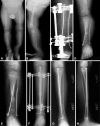
(A) Anteroposterior (AP) and (B) lateral view radiographs of a 2-year-old boy with neurofibromatosis and CPT (Type I) that had not been previously treated are shown. (C) AP view radiograph shows split of the proximal segment with invagination of the distal segment, rodding of tibia from medial malleolus and of fibula through lateral malleolus, and application of Ilizarov external fixator. (D) AP view radiograph obtained immediately after external fixator removed shows union of the CPT. (E) AP view radiograph shows refracture of the tibia 1 year later. (F) AP view radiograph shows retreatment with an Ilizarov device and rerodding with Fassier-Duval telescopic IM nail locked in both proximal and distal epiphysis. (G, H) Tibia remains healed 2 years later with good remodeling. The telescopic nail has extended with growth.

References

    1. {'text': '', 'ref_index': 1, 'ids': [{'type': 'PubMed', 'value': '819445', 'is_inner': True, 'url': 'https://pubmed.ncbi.nlm.nih.gov/819445/'}]}
    2. Andersen KS. Congenital pseudarthrosis of the leg. Late results. J Bone Joint Surg Am. 1976;58:657–662. - PubMed
    1. {'text': '', 'ref_index': 1, 'ids': [{'type': 'DOI', 'value': '10.1007/BF00387333', 'is_inner': False, 'url': 'https://doi.org/10.1007/bf00387333'}, {'type': 'PubMed', 'value': '6150696', 'is_inner': True, 'url': 'https://pubmed.ncbi.nlm.nih.gov/6150696/'}]}
    2. Blauth M, Harms D, Schmidt D, Blauth W. Light- and electron-microscopic studies in congenital pseudarthrosis. Arch Orthop Trauma Surg. 1984;103:269–277. - PubMed
    1. {'text': '', 'ref_index': 1, 'ids': [{'type': 'DOI', 'value': '10.1097/00004694-199709000-00019', 'is_inner': False, 'url': 'https://doi.org/10.1097/00004694-199709000-00019'}, {'type': 'PubMed', 'value': '9592010', 'is_inner': True, 'url': 'https://pubmed.ncbi.nlm.nih.gov/9592010/'}]}
    2. Boero S, Catagni M, Donzelli O, Facchini R, Frediani PV. Congenital pseudarthrosis of the tibia associated with neurofibromatosis-1: treatment with Ilizarov’s device. J Pediatr Orthop. 1997;17:675–684. - PubMed
    1. {'text': '', 'ref_index': 1, 'ids': [{'type': 'PubMed', 'value': '7083685', 'is_inner': True, 'url': 'https://pubmed.ncbi.nlm.nih.gov/7083685/'}]}
    2. Boyd HB. Pathology and natural history of congenital pseudarthrosis of the tibia. Clin Orthop Relat Res. 1982;166:5–13. - PubMed
    1. {'text': '', 'ref_index': 1, 'ids': [{'type': 'PubMed', 'value': '13610959', 'is_inner': True, 'url': 'https://pubmed.ncbi.nlm.nih.gov/13610959/'}]}
    2. Boyd HB, Sage FP. Congenital pseudarthrosis of the tibia. J Bone Joint Surg Am. 1958;40:1245–1270. - PubMed