Review
. 2008 Oct 27:6:113.
doi: 10.1186/1477-7819-6-113.

Intravascular leiomyosarcoma of the brachiocephalic region -- report of an unusual tumour localisation: case report and review of the literature

Affiliations
Review

Intravascular leiomyosarcoma of the brachiocephalic region -- report of an unusual tumour localisation: case report and review of the literature

Daniel-Johannes Tilkorn et al. World J Surg Oncol. .

Abstract

Background: Intravascular leiomyosarcoma is a rare tumour entity originating from venous vessel structures and most frequently affecting the inferior vena cava.

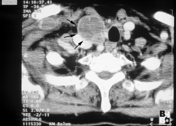
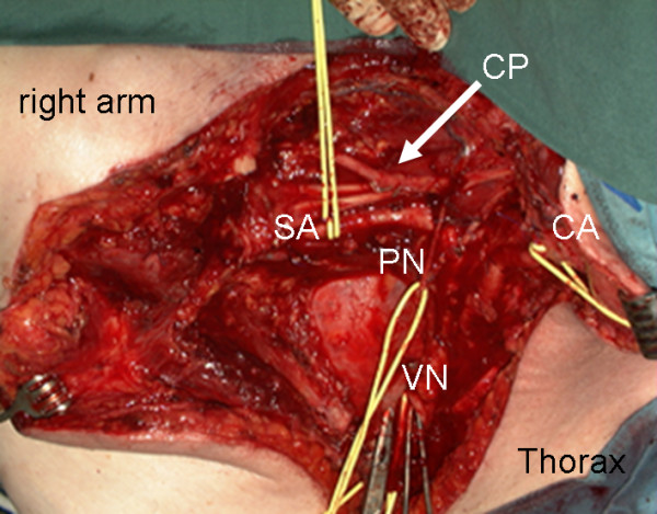
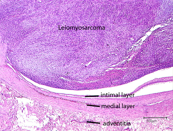
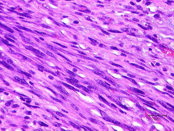
Case presentation: A 69-year old patient presented with a biopsy proven leiomyosarcoma of the right supraclavicular region. Tumour resection and histological assessment verified the intravascular tumour origin arising from the internal jugular vein and extending into the surrounding soft tissue.

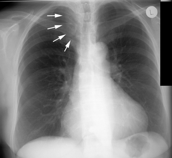
Conclusion: In the presence of a biopsy proven diagnosis of leiomyosarcoma the rare condition of an intravascular tumour origin has to be considered even without signs of venous stases. This may result in an altered surgical strategy. Microthrombembolism and pulmonary metastases may complicate the course of the disease.


Figures

Figure 1
Figure 1
CT scan of the neck and upper medastinum: Confirmation of a soft tissue tumour (→) 4 cm in size. Expansive tumour growth displaced the trachea to the left and compressed the adjacent vessels.
Figure 2
Figure 2
Preoperative chest x-ray displayed a mediastinal enlargement towards the right (→).
Figure 3
Figure 3
Surgical situs: a vessel loop was placed around the subclavian artery (SA), the carotic artery (CA), the right vagus nerve (VN) and the phrenic nerve (PN). CP indicates the cervical plexus; Clamps were placed on the stumps of the cut superior vena cava. The retractor on the left edge held back the pectoralis major muscle, in the center the exposed lung apex is visible.
Figure 4
Figure 4
Intraluminal tumour growth of a Leiomyosarcoma originating from the subclavian vein (H&E-staining).
Figure 5
Figure 5
"Cigar shaped" configurations of tumor cell nuclei of a leiomyosarcoma with nuclear atypia (H&E staining).

References

    1. Shimoda H, Oka K, Otani S, Hakozaki H, Yoshimura T, Okazaki H, Nishida S, Tomita S, Oka T, Kawasaki T, Mori N. Vascular leiomyosarcoma arising from the inferior vena cava diagnosed by intraluminal biopsy. Virchows Arch. 1998;433:97–100. doi: 10.1007/s004280050223. - DOI - PubMed
    1. Weiss SWGJ. Leiomysarcoma. In: Weiss SW, Goldblum JR (Hrsg), editor. Enzinger and Weiss's soft tissue tumors. 4. Mosby, StLouis Baltimore Berlin; 2001. pp. 727–748.
    1. Varela-Duran J, Oliva H, Rosai J. Vascular leiomyosarcoma: the malignant counterpart of vascular leiomyoma. Cancer. 1979;44:1684–1691. doi: 10.1002/1097-0142(197911)44:5<1684::AID-CNCR2820440523>3.0.CO;2-I. - DOI - PubMed
    1. Perl L. Ein Fall vom Sarkom der Vena cava inferior. Virchows Arch. 1871;53:378–385. doi: 10.1007/BF01957198. - DOI
    1. Subramaniam MM, Martinez-Rodriguez M, Navarro S, Rosaleny JG, Bosch AL. Primary intravascular myxoid leiomyosarcoma of the femoral vein presenting clinically as deep vein thrombosis: a case report. Virchows Arch. 2007;450:235–237. doi: 10.1007/s00428-006-0322-2. - DOI - PubMed

MeSH terms

LinkOut - more resources