. 2008 Oct 27:5:129.
doi: 10.1186/1743-422X-5-129.

Adaptation to cell culture induces functional differences in measles virus proteins

Affiliations

Adaptation to cell culture induces functional differences in measles virus proteins

Bettina Bankamp et al. Virol J. .

Abstract

Background: Live, attenuated measles virus (MeV) vaccine strains were generated by adaptation to cell culture. The genetic basis for the attenuation of the vaccine strains is unknown. We previously reported that adaptation of a pathogenic, wild-type MeV to Vero cells or primary chicken embryo fibroblasts (CEFs) resulted in a loss of pathogenicity in rhesus macaques. The CEF-adapted virus (D-CEF) contained single amino acid changes in the C and matrix (M) proteins and two substitutions in the shared amino terminal domain of the phosphoprotein (P) and V protein. The Vero-adapted virus (D-VI) had a mutation in the cytoplasmic tail of the hemagglutinin (H) protein.

Results: In vitro assays were used to test the functions of the wild-type and mutant proteins. The substitution in the C protein of D-CEF decreased its ability to inhibit mini-genome replication, while the wild-type and mutant M proteins inhibited replication to the same extent. The substitution in the cytoplasmic tail of the D-VI H protein resulted in reduced fusion in a quantitative fusion assay. Co-expression of M proteins with wild-type fusion and H proteins decreased fusion activity, but the mutation in the M protein of D-CEF did not affect this function. Both mutations in the P and V proteins of D-CEF reduced the ability of these proteins to inhibit type I and II interferon signaling.

Conclusion: Adaptation of a wild-type MeV to cell culture selected for genetic changes that caused measurable functional differences in viral proteins.


Figures

Figure 1
Figure 1
Expression of proteins derived from D87-wt, D-VI and D-CEF. (A, B, C) A549 cells were infected with vTF7-3 and transfected with the indicated pTM1-derived plasmids. (D) Vero cells were transfected with the indicated pCAGGS-derived plasmids. In all cases, proteins were labeled with 35S-methionine, precipitated with protein-specific antisera and separated by SDS-PAGE. Molecular mass markers (kDa) are shown on the left in each panel, the positions of proteins are indicated on the right.
Figure 2
Figure 2
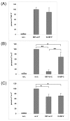
Effect of mutations in the P, C and V proteins of MeV on mini-genome replication. (A) CV-1 cells were infected with MVAT7 and transfected with pMV107(-)CAT, pTM1-D87-wt N, pTM1-D87-wt L and the indicated plasmids expressing P proteins. (B, C) CV-1 cells were infected with MVAT7 and transfected with pMV107(-)CAT, pTM1-D87-wt N, pTM1-D87-wt P, pTM1-D87-wt L and 1 μg of the indicated plasmids. For the negative controls, pTM1-D87-wt L was omitted. The amount of transfected DNA was kept constant through the addition of pTM1 vector. CAT protein production in cytoplasmic extracts of quadruplicate samples was measured by ELISA. The amount of CAT protein measured in the presence of pTM1-D87-wt P and absence of C- or V-expressing plasmids was set to 100% in each panel. Each panel shows the average of three independent experiments. Error bars denote one standard deviation. (*: P ≤ 0.01, **: P ≤ 0.001)
Figure 3
Figure 3
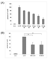
The M protein of MeV inhibits mini-genome replication. (A) CV-1 cells were infected with MVAT7 and transfected with pMV107(-)CAT, pTM1-D87-wt N, pTM1-D87-wt P, pTM1-D87-wt L and increasing amounts of pTM1-D87-wt M. (B) CV-1 cells were infected with MVAT7 and transfected with pMV107(-)CAT, pTM1-D87-wt N, pTM1-D87-wt P, pTM1-D87-wt L and 2 μg of the indicated plasmids. Transfections and ELISA were performed as described in the legend to figure 1. (*: P ≤ 0.01)
Figure 4
Figure 4
Effect of mutations in the H and M proteins of MeV on quantitative fusion. (A) fusion produced by H and F proteins, (B) inhibition of fusion by co-expressed M protein. Vero/hSLAM cells were infected with MVAT7 and transfected with the indicated plasmids. The amount of transfected DNA was kept constant through the addition of pTM1 vector. β-galactosidase protein production in cytoplasmic extracts of quadruplicate samples was measured as described in the Methods section. The amount of β-galactosidase protein measured in the wells transfected with pTM1-D87-wt F and pTM1-D87-wt H was set to 100% in each panel. Each panel shows the average of three independent experiments. Error bars denote one standard deviation. In panels (A) and (B) F indicates D87-wt F, in panel (B), H indicates D87-wt H. (**: P ≤ 0.001)
Figure 5
Figure 5
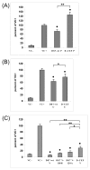
Cell culture adaptation affects the inhibition of IFN-β signaling by the P, C, and V proteins of MeV. Vero cells were transfected with a plasmid constitutively expressing renilla luciferase, a plasmid expressing firefly luciferase under the control of an IFN-α/β-responsive promoter and the plasmids as indicated in the three panels. Forty-eight hours after transfection, cells were stimulated with IFN-β for six hours, lysed and tested for luciferase activity. VC-indicates cells transfected with pCAGGS empty vector but not stimulated while VC+ indicates cells transfected with pCAGGS empty vector and stimulated with IFN-β. Results are expressed as a ratio of firefly to renilla luciferase luminescence taken as a percentage of the luminescence obtained using IFN-stimulated, empty pCAGGS vector (VC+). (A) results of IFN-β signaling assay with P proteins from D87-wt and D-CEF, (B, C) inhibition of IFN-β signaling by the C and V proteins, respectively. The data shown are an average of three experiments done with triplicate samples. Error bars denote one standard deviation. Bars marked with a are significantly different from VC+ with a P ≤ 0.05. (*: P ≤ 0.01, **: P ≤ 0.001)
Figure 6
Figure 6
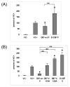
Cell culture adaptation affects the inhibition of IFN-γ signaling by the P and V proteins of MeV. Experiments were performed as described in the legend to figure 5, except that a reporter plasmid expressing firefly luciferase under the control of an IFN-γ-responsive promoter and IFN-γ were used for stimulation. (A) results of IFN-γ signaling assays with P proteins from D87-wt and D-CEF, (B) inhibition of IFN-γ signaling by the V proteins. Bars marked with a are significantly different from VC+ with a P ≤ 0.05. (**: P ≤ 0.001)
Figure 7
Figure 7
Location of IFN-inhibiting domains in the P and V proteins of MeV. P indicates P protein, V indicates V protein, NTD indicates shared amino terminal domain, and CTD indicates unique carboxyl terminal domain. The black bars denote the domain from amino acids 110–130 in the NTD. Positions of important amino acids are marked.

References

    1. Hilleman MR, Buynak EB, Weibel RE, Stokes J, Jr, Whitman JE, Jr, Leagus MB. Development and evaluation of the Moraten measles virus vaccine. Jama. 1968;206:587–590. doi: 10.1001/jama.206.3.587. - DOI - PubMed
    1. Ikic D, Juzbasic M, Beck M, Hrabar A, Cimbur-Schreiber T. Attenuation and characterisation of Edmonston-Zagreb measles virus. Ann Immunol Hung. 1972;16:175–181. - PubMed
    1. Sasaki K. Studies on the modification of the live AIK measles vaccine. I. Adaptation of the further attenuated AIK measles virus (the strain AIK-L33) to chick embryo cells. Kitasato Arch Exp Med. 1974;47:1–12. - PubMed
    1. Schwarz AJ. Immunization against Measles: Development and Evaluation of a Highly Attenuated Live Measles Vaccine. Ann Paediatr. 1964;202:241–252. - PubMed
    1. Parks CL, Lerch RA, Walpita P, Wang HP, Sidhu MS, Udem SA. Analysis of the noncoding regions of measles virus strains in the Edmonston vaccine lineage. J Virol. 2001;75:921–933. doi: 10.1128/JVI.75.2.921-933.2001. - DOI - PMC - PubMed

Publication types

LinkOut - more resources