

Proximal colon distension induces Fos expression in oxytocin-, vasopressin-, CRF- and catecholamines-containing neurons in rat brain
- PMID: 18955037
- PMCID: PMC3210201
- DOI: 10.1016/j.brainres.2008.09.094
Proximal colon distension induces Fos expression in oxytocin-, vasopressin-, CRF- and catecholamines-containing neurons in rat brain
Abstract
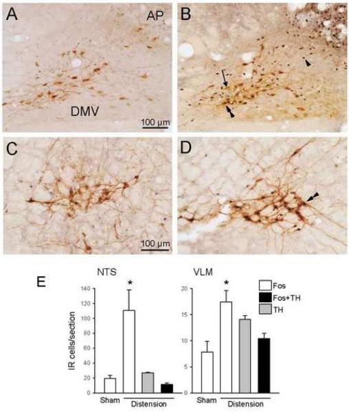
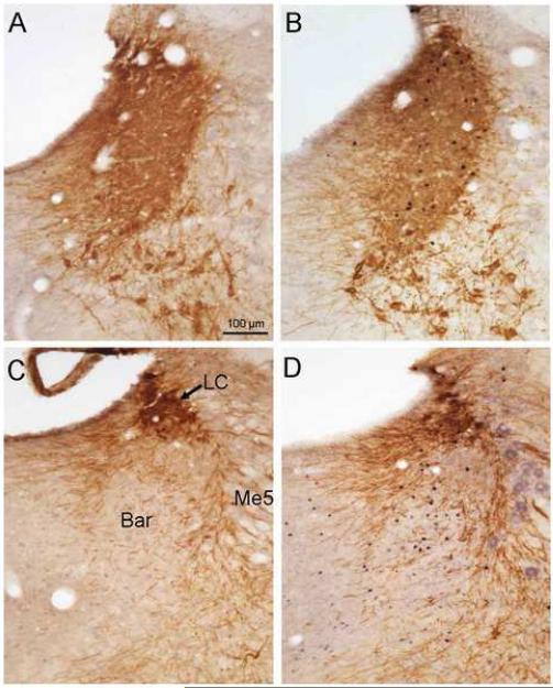
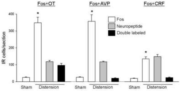
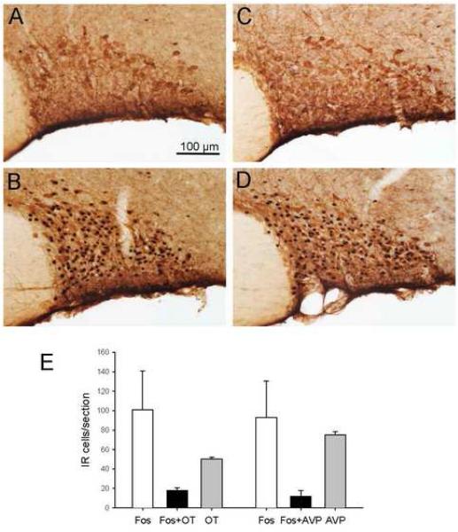
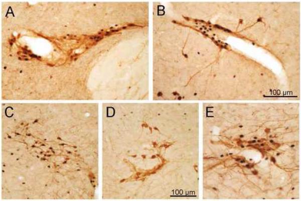
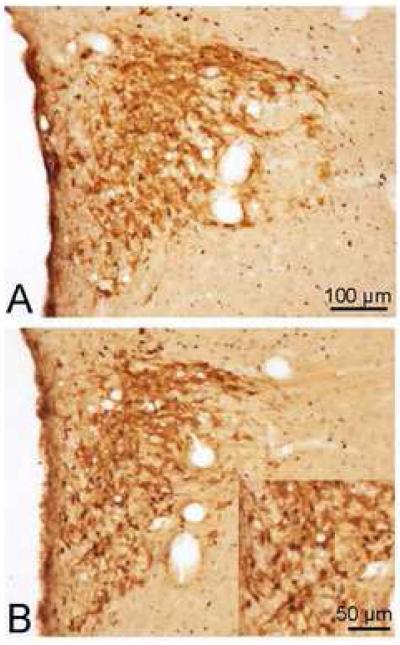
Little is known about the chemical coding of the brain neuronal circuitry activated by nociceptive signals of visceral origin. We characterized brain nuclei activated during isovolumetric phasic distension of the proximal colon (10 ml, 30 s on/off for 10 min) in conscious male rats, using Fos as a marker of neuronal activation and dual immunohistochemistry to visualize co-localization of Fos expression and oxytocin (OT), arginine-vasopressin (AVP), corticotrophin-releasing factor (CRF) or tyrosine hydroxylase (TH). Proximal colon distension, compared with sham distension, induced a robust increase in Fos-like immunoreactive (IR) neurons in the paraventricular nucleus (PVN), supraoptic nucleus (SON) and accessory neurosecretory nuclei of the hypothalamus, nucleus of the solitary tract (NTS) and ventrolateral medulla (VLM), and to a lower extent, in the locus coeruleus (LC) and Barrington nucleus. Fos-IR neurons in the PVN after colon distension were identified in 81% of OT-IR, 18% AVP-IR and 16% CRF-IR neurons, while in the SON it represented 36% of OT-IR and 16% AVP-IR. Catecholaminergic cell groups in the pons (LC) and medulla (VLM, NTS) were also activated by proximal colon distension. Of the TH-IR neurons in VLM and NTS, 74% and 42% respectively were double labeled. These results indicate that colon distension stimulates OT-, AVP- and CRF-containing hypothalamic neurons, likely involved in the integration of colonic sensory information to modulate autonomic outflow and pain-related responses. Activation of medullary catecholaminergic centers might reflect the afferent and efferent limbs of the functional responses associated to visceral pain.
Figures

Similar articles
-
Histamine stimulates c-fos expression in hypothalamic vasopressin-, oxytocin-, and corticotropin-releasing hormone-containing neurons.Endocrinology. 1994 Jan;134(1):482-91. doi: 10.1210/endo.134.1.8275963. Endocrinology. 1994. PMID: 8275963
-
Oxytocin and vasopressin involved in restraint water-immersion stress mediated by oxytocin receptor and vasopressin 1b receptor in rat brain.PLoS One. 2011;6(8):e23362. doi: 10.1371/journal.pone.0023362. Epub 2011 Aug 17. PLoS One. 2011. PMID: 21858088 Free PMC article.
-
C-fos mRNA pattern and corticotropin-releasing factor neuronal activity throughout the brain of rats injected centrally with a prostaglandin of E2 type.J Neuroimmunol. 1996 Nov;70(2):163-79. doi: 10.1016/s0165-5728(96)00114-2. J Neuroimmunol. 1996. PMID: 8898725
-
Abdominal surgery activates nesfatin-1 immunoreactive brain nuclei in rats.Peptides. 2010 Feb;31(2):263-70. doi: 10.1016/j.peptides.2009.11.015. Epub 2009 Nov 26. Peptides. 2010. PMID: 19944727 Free PMC article.
-
The role of catecholaminergic neurons in the hypothalamus and medullary visceral zone in response to restraint water-immersion stress in rats.J Physiol Sci. 2011 Jan;61(1):37-45. doi: 10.1007/s12576-010-0119-6. Epub 2010 Dec 16. J Physiol Sci. 2011. PMID: 21161464 Free PMC article.
Cited by
-
Pattern of Fos expression in the brain induced by selective activation of somatostatin receptor 2 in rats.Brain Res. 2010 Sep 10;1351:150-164. doi: 10.1016/j.brainres.2010.07.024. Epub 2010 Jul 15. Brain Res. 2010. PMID: 20637739 Free PMC article.
-
The expression of corticotropin-releasing factor in the central nucleus of the amygdala, induced by colorectal distension, is attenuated by general anesthesia.J Korean Med Sci. 2010 Nov;25(11):1646-51. doi: 10.3346/jkms.2010.25.11.1646. Epub 2010 Oct 26. J Korean Med Sci. 2010. PMID: 21060755 Free PMC article.
-
Functional disruption of cortical cingulate activity attenuates visceral hypersensitivity and anxiety induced by acute experimental colitis.Sci Rep. 2021 Jan 22;11(1):2103. doi: 10.1038/s41598-021-81256-x. Sci Rep. 2021. PMID: 33483524 Free PMC article.
-
Corticotrophin-releasing factor 1 activation in the central amygdale and visceral hyperalgesia.Neurogastroenterol Motil. 2015 Jan;27(1):1-6. doi: 10.1111/nmo.12495. Neurogastroenterol Motil. 2015. PMID: 25557223 Free PMC article. Review.
-
Brain corticotropin-releasing factor signaling: Involvement in acute stress-induced visceral analgesia in male rats.Neurogastroenterol Motil. 2019 Feb;31(2):e13489. doi: 10.1111/nmo.13489. Epub 2018 Oct 9. Neurogastroenterol Motil. 2019. PMID: 30298965 Free PMC article.
References
-
- Al Chaer ED, Kawasaki M, Pasricha PJ. A new model of chronic visceral hypersensitivity in adult rats induced by colon irritation during postnatal development. Gastroenterology. 2000;119:1276–1285. - PubMed
-
- Amico JA, Mantella RC, Vollmer RR, Li X. Anxiety and stress responses in female oxytocin deficient mice. J. Neuroendocrinol. 2004;16:319–324. - PubMed
-
- Azpiroz F, Malagelada JR. Intestinal control of gastric tone. Am. J. Physiol. 1985;249:G501–G509. - PubMed
-
- Benarroch EE. Pain-autonomic interactions. Neurol. Sci. 2006;27(Suppl 2):S130–S133. - PubMed
-
- Birder LA, de Groat WC. Induction of c-fos expression in spinal neurons by nociceptive and nonnociceptive stimulation of LUT. Am. J. Physiol. 1993;265:R326–R333. - PubMed
Publication types
MeSH terms
Substances
Grants and funding
LinkOut - more resources
Full Text Sources
Medical
Miscellaneous
