. 2008 Dec 19;283(51):35644-51.
doi: 10.1074/jbc.M806351200. Epub 2008 Oct 27.

Residue 17 of sauvagine cross-links to the first transmembrane domain of corticotropin-releasing factor receptor 1 (CRFR1)

Affiliations

Residue 17 of sauvagine cross-links to the first transmembrane domain of corticotropin-releasing factor receptor 1 (CRFR1)

Iman Assil-Kishawi et al. J Biol Chem. .

Abstract

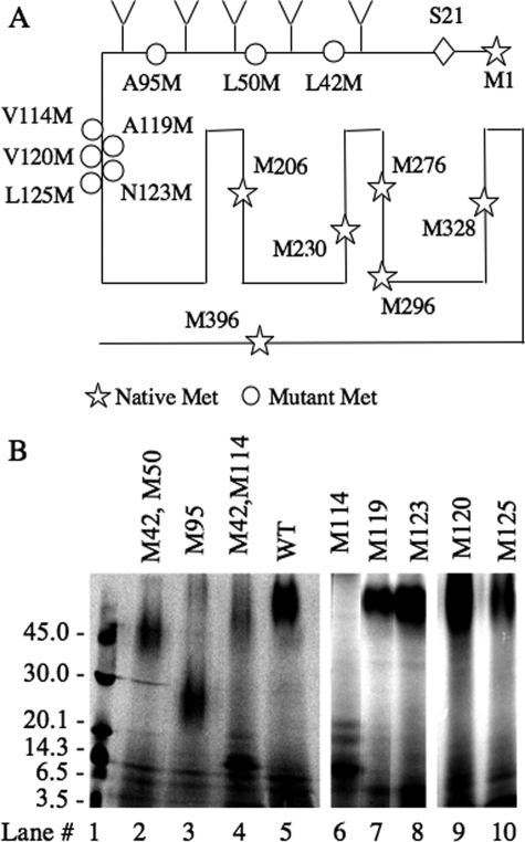
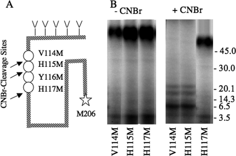
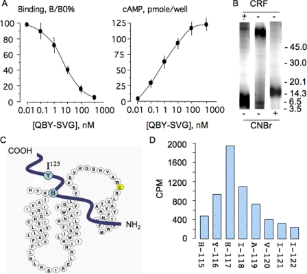
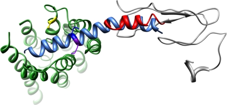
Corticotropin-releasing factor receptor 1 (CRFR1) mediates the physiological actions of corticotropin-releasing factor in the anterior pituitary gland and the central nervous system. Using chemical cross-linking we have previously reported that residue 16 of sauvagine (SVG) is in a close proximity to the second extracellular loop of CRFR1. Here we introduced p-benzoylphenylalanine (Bpa) at position 17 of a sauvagine analog, [Tyr0, Gln1, Bpa17]SVG, to covalently label CRFR1 and characterize the cross-linking site. Using a combination of receptor mutagenesis, peptide mapping, and N-terminal sequencing, we identified His117 within the first transmembrane domain (TM1) of CRFR1 as the cross-linking site for Bpa17 of 125I-[Tyr0, Gln1, Bpa17]SVG. These data indicate that, within the SVG-CRFR1 complex, residue 17 of the ligand lies within a 9 angstroms distance from residue 117 of the TM1 of CRFR1. The molecular proximity between residue 17 of the ligand and TM1 of CRFR1 described here and between residue 16 of the ligand and the CRFR1 second extracellular loop described previously provides useful molecular constraints for modeling ligand-receptor interaction in mammalian cells expressing CRFR1.


Figures

FIGURE 1.
FIGURE 1.
Properties of the Bpa-substituted sauvagine, [Tyr0, Gln1, Bpa17]SVG (YQB-SVG). A, binding and signaling properties. Intact LLCPK-1 cells expressing mCRFR1 were incubated with 125I-YQ-SVG (∼100,000 cpm) in the presence of increasing concentrations of YQB-SVG or YQ-SVG for 2 h at 18 C; the cells were rinsed (three times) with binding buffer; the cells were lysed with 1 m NaOH (0.25 ml × 3), and the amount of bound radioligand was determined by counting in a γ-counter. The data (B/B0%) are calculated from the means ± S.D. of triplicates. Stimulation of cAMP accumulation by YQB-SVG and YQ-SVG was performed in LLCPK-1 cells expressing CRFR1. The cells were challenged with increasing concentration of the agonist for 20 min in presence of 2 mm 3-isobutyl-1-methylxanthene, supernatant was removed, and intracellular cAMP was extracted by lysing the cells with 100 mm HCl and measured by specific radioimmunoassay. The data are the means ± S.D. of triplicates. B, photoaffinity cross-linking of 125I-YQ-SVG to LLCPK-1 cells stably expressing mCRFR1. The cells were incubated with 125I-YQB-SVG (200,000 cpm/well) in presence of increasing concentrations of YQB-SVG for 2 h at 18 C; the cells were then rinsed with binding buffer and exposed to UV light. The receptor-ligand complexes were extracted with SDS sample buffer and loaded on 5–20% SDS-PAGE, followed by autoradiography.
FIGURE 2.
FIGURE 2.
Mapping of the cross-linked sites using CNBr cleavage of CRFR1 bearing Met substitution. A, the locations of native methionine residues in the CRFR1 backbone are depicted with stars, and the sites of the methionine substitutions are illustrated with circles; Ser24 is predicted to be the first residue of the mature protein (40, 41). B, CRFR1 or CRFR1 with methionine residues substitutions are expressed in COS-7 cells. Binding and cross-linking of 125I-YQB-SVG to the different CRFR1 mutants, expressed in COS-7 cells, was performed to as in Fig. 1B. The cell lysates were then cleaved with CNBr. The cleaved products were lyophilized, resuspended in sample buffer, and analyzed on Tricine-PAGE followed by autoradiography. Lane 1 shows the elution profile of the molecular mass markers. WT, wild type.
FIGURE 3.
FIGURE 3.
Cross-linking efficiency of CRFR1 mutants. The different mutants were expressed in COS-7 cells; binding and cross-linking to 125I-YQB-SVG was performed as in Fig. 1B. Similar amounts of bound receptor-ligand complex (1200 cpm) were analyzed on SDS-PAGE followed by autoradiography using phosphorimaging (A). In B, cross-linking efficiency was calculated as the ratios of the receptor band of the different receptor mutants divided by the receptor band in the wild type. WT, wild type.
FIGURE 4.
FIGURE 4.
Characterization of cross-linked CNBr-cleaved fragments of V114M, H115M and H117M CRFR1 mutants. A, scheme showing the location of the three CRFR1 methionine (M) mutations, the endogenous methionine at position 206, and the potential cross-linked site. B, the different CRFR1 mutants, expressed in COS-7 cells were cross-linked to 125I-YQB-SVG as in Fig. 1B and then analyzed on Tricine-PAGE before (-CNBr) and after (+CNBr) CNBr cleavage. The cleaved products were lyophilized, resuspended in sample buffer, and analyzed on Tricine-PAGE followed by autoradiography. The positions of the molecular mass markers are shown on the right side of the gel. The Y116M receptor mutant was not functional, and cross-linking could not be performed.
FIGURE 5.
FIGURE 5.
A, competition binding with [Gln1, Bpa17, Tyr23]SVG (QBY-SVG) against 125I-QBY-SVG and stimulation of cAMP accumulation in LLCPK-1 cells stably expressing CRFR1; the experimental conditions are similar to those in Fig. 1A. The data are the means ± S.D. of triplicates. B, photoaffinity cross-linking of 125I-QBY-SVG to COS-7 cells expressing the Met114 CRFR1 mutant in presence and absence of excess ovine CRF (1 μm). CNBr cleavage of the photo-cross-linked materials were resolved on Tricine-PAGE. C, scheme showing the relative position of Tyr23 and Bpa17 in the QBY-SVG analog and the cross-linked site His117 in CRFR1. D, radiosequencing of cross-linked receptor-ligand fragment purified from the 16-kDa band depicted in B and sequenced using Edman degradation; at each cycle the radioactivity released was quantified in a γ-counter for 5 min; ∼60% of radioactivity submitted for sequencing was recovered in the fractions.
FIGURE 6.
FIGURE 6.
Molecular model of sauvagine binding to CRF1R. The structure of sauvagine, as determined by homology with human urocortin-1, is shown in aqua, with Lys16 and Met17 denoted in dark blue and purple, respectively. The orientation of sauvagine to CRFR1 was guided by the NMR structure of the N terminus of CRFR2 (gray) bound to the C-terminal portion of astressin (red). The 19 residues missing between the CRF2R N terminus as determined by NMR and the beginning of TM1 is denoted by a dashed blue line. Likewise, the EC2 consisting of KLYYDNEKCWFGKRPGVYT is denoted as an orange dashed line, with Cys258 forming a disulfide bond with Cys188 at the extracellular end of TM3 (shown in yellow).

References

    1. Vale, W., Spiess, J., Rivier, C., and Rivier, J. (1981) Science 213 1394-1397 - PubMed
    1. Chang, C. P., Pearse, R. V., II, O'Connell, S., and Rosenfeld, M. G. (1993) Neuron 11 1187-1195 - PubMed
    1. Chen, R., Lewis, K. A., Perrin, M. H., and Vale, W. W. (1993) Proc. Natl. Acad. Sci. U. S. A. 90 8967-8971 - PMC - PubMed
    1. Kishimoto, T., Pearse, R. V., II, Lin, C. R., and Rosenfeld, M. G. (1995) Proc. Natl. Acad. Sci. U. S. A. 92 1108-1112 - PMC - PubMed
    1. Lovenberg, T. W., Liaw, C. W., Grigoriadis, D. E., Clevenger, W., Chalmers, D. T., De Souza, E. B., and Oltersdorf, T. (1995) Proc. Natl. Acad. Sci. U. S. A. 92 836-840 - PMC - PubMed

Publication types

MeSH terms

LinkOut - more resources