. 2009 Jan;29(2):471-82.
doi: 10.1128/MCB.01352-08. Epub 2008 Oct 27.

Functional dissection of human and mouse POT1 proteins

Affiliations

Functional dissection of human and mouse POT1 proteins

Wilhelm Palm et al. Mol Cell Biol. 2009 Jan.

Abstract

The single-stranded telomeric DNA binding protein POT1 protects mammalian chromosome ends from the ATR-dependent DNA damage response, regulates telomerase-mediated telomere extension, and limits 5'-end resection at telomere termini. Whereas most mammals have a single POT1 gene, mice have two POT1 proteins that are functionally distinct. POT1a represses the DNA damage response, and POT1b controls 5'-end resection. In contrast, as we report here, POT1a and POT1b do not differ in their ability to repress telomere recombination. By swapping domains, we show that the DNA binding domain of POT1a specifies its ability to repress the DNA damage response. However, no differences were detected in the in vitro DNA binding features of POT1a and POT1b. In contrast to the repression of ATR signaling by POT1a, the ability of POT1b to control 5'-end resection was found to require two regions in the C terminus, one corresponding to the TPP1 binding domain and a second representing a new domain located between amino acids (aa) 300 and 350. Interestingly, the DNA binding domain of human POT1 can replace that of POT1a to repress ATR signaling, and the POT1b region from aa 300 to 350 required for the regulation of the telomere terminus is functionally conserved in human POT1. Thus, human POT1 combines the features of POT1a and POT1b.


Figures
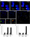
FIG. 1.
FIG. 1.
POT1a and -b repress HDR in parallel with Ku70. (A) Examples of T-SCE events in POT1 DKO cells deficient for Ku70. CO-FISH analysis of POT1aS/F POT1bS/F Ku70−/− MEFs was performed 92 h after treatment with adenoviral Cre. Fluorescence signals of the TelG and TelC probes are depicted separately and merged. The enlargements depict chromosomes exhibiting T-SCEs. (B) Comparable induction of T-SCEs upon the simultaneous removal of POT1a and POT1b or the removal of TRF2 in Ku70−/− cells (POT1 DKO Ku70−/−, P = 0.006 by Student's t test). Shown is the quantification of three independent CO-FISH experiments; at least 6,000 chromosome ends were analyzed for the POT1aS/F POT1bS/F cell lines and at least 4,000 for the TRF2FLOX/− Ku70−/− cell lines. The increase in T-SCE frequency after POT1a/b deletion in Ku70-proficient cells is not statistically significant (P = 0.184). (C) Both POT1a and POT1b alone are capable of suppressing T-SCEs in Ku70−/− MEFs. Shown is the quantification of three independent CO-FISH experiments. At least 1,000 chromosome ends were analyzed for each cell line in each experiment. Error bars in panels B and C indicate standard deviations.
FIG. 2.
FIG. 2.
Binding of POT1a and -b to 3′ and 5′ telomeric sites. (A and B) Gel-shift reactions with the probes indicated below the gels and increasing amounts of POT1a and POT1b. (A) Binding of POT1a and -b to the probes a, a3, and a3′. The red letters indicate nucleotide changes that interfere with POT1 binding to the 5′ site. The minimal binding sites are highlighted by the red boxes. Protein amounts in each binding reaction are indicated above the lanes in the left panel, and the same amounts were used in other panels. (B) POT1a and -b bind to the telomeric site at a 5′ end with diminished affinity. The mutations in primer a5 block POT1a and POT1b from binding to the 3′ end of the probe. Probes with no POT1 recognition site are not bound by POT1a or -b. The gel shifts shown are representatives of three independent experiments. (C) Summary of the Kd values (nM) of POT1a and POT1b in gel-shift experiments with the indicated probes. Values derived from three or more experiments are given as averages with standard deviations. Where probes were tested only twice, both values are given. All experiments were done with POT1a and -b in parallel, and Kd values were derived from quantitative analysis of the phosphorimager data as shown in panels A and B.
FIG. 3.
FIG. 3.
Both POT1a and -b bind equally to telomeric ds-ss junctions. (A to C) Binding of POT1a and -b to an ss telomeric site near a ds-ss junction. The probes were incubated with increasing POT1a and -b concentrations (0.156 to 40 nM) for 30 min. Sequences of the ds-ss regions of the probes are indicated below the gels; their sequences are given in Fig. S1 in the supplemental material. All probes have a sequence change in the 3′ end blocking binding to the 3′ end. (A) Diminished binding to mutant telomere [Telo(mut)] and telomere 4 (Telo4) (shown below the gels) in which the G residue 5′ of the site is altered or base paired. (B) Diminished binding of POT1a and -b to additional probes with ds DNA next to the binding site occluding the G residues 5′ of the binding site. (C) No diminished binding to probes with available G residues 5′ of the binding site. The gel shifts shown are representatives of three independent experiments. (D) Summary of the Kd values (nM) of POT1a and POT1b in gel-shift experiments with the indicated probes. Values derived from three or more experiments are given as averages with standard deviations. Where probes were tested only twice, both values are given. All experiments were done with POT1a and -b in parallel, and Kd values were derived from quantitative analysis of phosphorimager data as shown in panels A to C.
FIG. 4.
FIG. 4.
Structure, expression, and localization of POT1 chimeras. (A) POT1a, POT1b, and human POT1 (hPOT1) were divided into N- and C-terminal parts between the second OB fold of the DNA binding domain and the TPP1 interaction domain and fused together according to the six possible permutations. In the schematic depiction, red corresponds to POT1a, green to POT1b, and blue to human POT1. For one set of POT1 chimeras (AB, AH, BA, BH, HA, and HB), the swapping of the domains takes place at aa 350 (POT1a and POT1b) or aa 344 (human POT1). The domain swap of a second set of chimeras (AB2, AH2, HA2, and HB2) takes place at the very end of the second OB fold at aa 301 (POT1a) and aa 299 (human POT1). (B) Immunoblot on cells expressing POT1a, POT1b, human POT1, or POT1 chimeras. MEFs conditionally targeted for POT1a and POT1b (POT1aS/F POT1bS/F) were infected with pWZL-N-myc-POT1 or vector (pWZL) and cell extracts prepared after hygromycin selection. (C) Immunoblot on cells expressing POT1b or POT1 chimeras and if indicated hTPP1. MEFs conditionally targeted for POT1b (POT1bS/F) were infected with pWZL-N-myc-POT1 or vector (pWZL) and selected with hygromycin. Cells were then infected with pLPC-N-flag-hTPP1 or vector (pLPC), and cell extracts prepared after puromycin selection. AB2′-expressing cells represent an independent infection of POT1bS/F MEFs with AB2. The asterisk indicates a nonspecific band. (D) IF on cells expressing POT1a, POT1b, human POT1, or chimeric POT1 proteins. hTPP1 was introduced into the POT1aS/F POT1bS/F MEFs depicted in panel B that were expressing POT1 constructs with the C terminus of human POT1; otherwise, cells were infected with empty vector (pLPC). Endogenous POT1a and POT1b were deleted with adenoviral Cre. Cells were analyzed by IF for the myc epitope tag of the POT1 variants (green) and TRF1 (red) and counterstained with DAPI (4′,6-diamidino-2-phenylindole; blue). Similar results were obtained for wild-type cells (data not shown), but the telomeric accumulation of ectopic POT1 variants in the presence of endogenous POT1 was lower overall. Antibodies used for the experiments depicted in panels B to D are 9E10 for myc, M2 for Flag, 644 for TRF1, 1151 for TPP1, and GTU88 for γ-tubulin (γ-tub).
FIG. 5.
FIG. 5.
Chimeric POT1 proteins suppress the growth defects of POT1a and POT1b DKO cells. Growth curves of MEFs conditionally targeted for both POT1a and POT1b (POT1aS/F POT1bS/F) expressing the different POT1 variants and, if indicated, hTPP1 after the deletion of endogenous POT1a and POT1b with adenoviral Cre. The growth curves depicted in panels A to D were acquired in parallel and in duplicate; the growth curves depicted in panels E to G were acquired in parallel and in triplicate. (H) Summary of the capability of human and mouse POT1 proteins and their chimeras to rescue cell proliferation in POT1a and POT1b DKO MEFs. ++, efficient suppression of growth defects (comparable to POT1a); +, partial suppression of growth defects; −, no suppression of growth defects.
FIG. 6.
FIG. 6.
The DNA binding domains of POT1a or human POT1 repress ATR signaling. (A) MEFs conditionally targeted for POT1a and POT1b (POT1aS/F POT1bS/F) expressing the different POT1 variants and, if indicated, hTPP1 were infected with adenoviral Cre. Four days after the application of Cre, the occurrence of TIFs was monitored by IF for γ-H2AX (green) and TRF1 (red), and counterstained with DAPI (4′,6-diamidino-2-phenylindole; blue). The growth curves depicted in Fig. 5E to G were obtained from the same set of Cre-infected cells. (B) Schematic representation of the POT1 chimeras that can repress ATR signaling. Chimeric proteins with the DNA binding domain of POT1a or human POT1 efficiently suppress TIF formation; the DNA binding domain of POT1b can do so but to a minor extent. The provenance of the C terminus is not critical in this regard, although POT1 variants with the human C terminus require the coexpression of hTPP1 for telomeric localization.
FIG. 7.
FIG. 7.
The POT1b C terminus is required to control 5′-end resection. (A) In-gel overhang assay of MEFs conditionally targeted for POT1b (POT1bS/F) expressing POT1b or POT1 chimeras. Cells were retrovirally transduced with the indicated POT1 variants in pWZL-N-myc or vector control (pWZL) and selected with hygromycin. Subsequently, cells were infected with pLPC-N-flag-hTPP1 or vector (pLPC) and selected with puromycin. POT1b was deleted using Hit&Run Cre, and overhang length was determined 6 days after the application of Cre. (B) Quantification of the overhang signals of the cells depicted in panel A. AB2′-expressing cells represent an independent infection of POT1bS/F MEFs with AB2.
FIG. 8.
FIG. 8.
Summary of the ability of the indicated POT1 chimeras to prevent the inappropriate resection of the telomeric 5′ end in the absence of POT1b. Results of the overhang assays are shown in Fig. 7 and in Fig. S5 in the supplemental material. For chimeras containing the C terminus of human POT1, hTPP1 was coexpressed in some experiments as indicated. +, repression of the increase in the overhang signal after the deletion of POT1b; −, minimal or no repression of the overhang signal; DN, no rescue of the overhang signal increase after the deletion of POT1b, combined with an increase in the overhang signal in cells containing POT1b. The asterisks indicate experiments in which human POT1 was not localized to telomeres due to the absence of hTPP1.

Similar articles

Cited by

References

    1. Baumann, P., and T. R. Cech. 2001. Pot1, the putative telomere end-binding protein in fission yeast and humans. Science 2921171-1175. - PubMed
    1. Celli, G., and T. de Lange. 2005. DNA processing not required for ATM-mediated telomere damage response after TRF2 deletion. Nat. Cell Biol. 7712-718. - PubMed
    1. Celli, G. B., E. Lazzerini Denchi, and T. de Lange. 2006. Ku70 stimulates fusion of dysfunctional telomeres yet protects chromosome ends from homologous recombination. Nat. Cell Biol. 8885-890. - PubMed
    1. Churikov, D., and C. M. Price. 2008. Pot1 and cell cycle progression cooperate in telomere length regulation. Nat. Struct. Mol. Biol. 1579-84. - PMC - PubMed
    1. de Lange, T. 2005. Shelterin: the protein complex that shapes and safeguards human telomeres. Genes Dev. 192100-2110. - PubMed

Publication types

MeSH terms

LinkOut - more resources