. 2008 Nov 3;183(3):401-8.
doi: 10.1083/jcb.200805107. Epub 2008 Oct 27.

Memo-RhoA-mDia1 signaling controls microtubules, the actin network, and adhesion site formation in migrating cells

Affiliations

Memo-RhoA-mDia1 signaling controls microtubules, the actin network, and adhesion site formation in migrating cells

Kossay Zaoui et al. J Cell Biol. .

Abstract

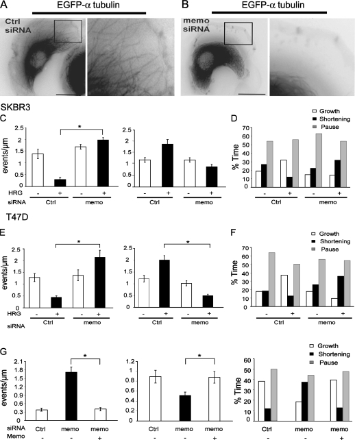
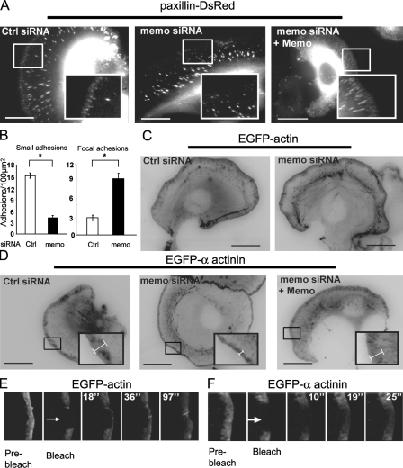
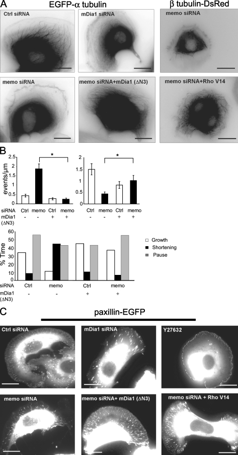
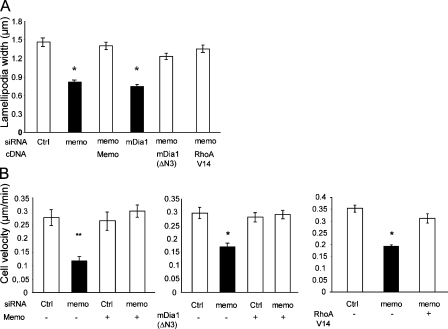
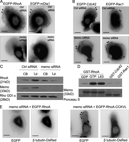
Actin assembly at the cell front drives membrane protrusion and initiates the cell migration cycle. Microtubules (MTs) extend within forward protrusions to sustain cell polarity and promote adhesion site turnover. Memo is an effector of the ErbB2 receptor tyrosine kinase involved in breast carcinoma cell migration. However, its mechanism of action remained unknown. We report in this study that Memo controls ErbB2-regulated MT dynamics by altering the transition frequency between MT growth and shortening phases. Moreover, although Memo-depleted cells can assemble the Rac1-dependent actin meshwork and form lamellipodia, they show defective localization of lamellipodial markers such as alpha-actinin-1 and a reduced number of short-lived adhesion sites underlying the advancing edge of migrating cells. Finally, we demonstrate that Memo is required for the localization of the RhoA guanosine triphosphatase and its effector mDia1 to the plasma membrane and that Memo-RhoA-mDia1 signaling coordinates the organization of the lamellipodial actin network, adhesion site formation, and MT outgrowth within the cell leading edge to sustain cell motility.


Figures

Figure 1.
Figure 1.
Memo is required for ErbB2-regulated MT dynamics. (A and B) SKBR3 cells expressing EGFP-tubulin and control (Ctrl; A) or Memo siRNA (B) were treated with 5 nM HRG for 10 min. Still images from Videos 1 and 2 (available at http://www.jcb.org/cgi/content/full/jcb.200805107/DC1) show limited MT outgrowth in Memo-depleted cells relative to control cells. Right panels show enlargement of boxed areas. (C–F) Individual MT plus ends were tracked over time after addition of HRG to SKBR3 (C and D) and T47D (E and F) cells, and dynamic instability was analyzed. Frequencies of rescues (right) and catastrophes (C and E, left) and the percentage of time MTs spent growing, shortening, or paused (D and F) were calculated. (G) Reexpression of Memo into HRG-treated Memo-depleted SKBR3 cells restores normal MT dynamics. Left, catastrophes; middle, rescues. Values are means ± SD from three independent experiments (30 MTs and nine cells). *, P < 0.05 by permutation analysis; n = 30. Bars, 10 μm.
Figure 2.
Figure 2.
Memo is involved in small adhesion site formation and α-actinin lamellipodial localization. (A) Still images from Videos 3 and 4 showing paxillin-DsRed–labeled adhesion sites in SKBR3 cells. Paxillin incorporates into two populations of adhesion sites with distinct localizations, sizes, and lifespan (left). Memo is required for the formation of small peripheral adhesion (middle vs. left). Reexpression of Memo restores small adhesion sites (right). (B) Number of small and large (focal) adhesions in control and Memo-depleted cells. Values are means per 100 μm2 ± SEM from three independent experiments (four cells and three regions per cell). *, P < 0.05 by Mann-Whitney. (C and D) Still images from Videos 5–8 showing EGFP-actin (C) or EGFP–α-actinin-1 (D) expression in control and Memo-depleted migrating cells. Width of α-actinin–labeled lamellipodia (brackets) is reduced in Memo-depleted cells (middle) and restored upon Memo reexpression (right). Actin labeling is unchanged. Insets show enlargements of leading edges. (E and F) EGFP-actin (E) or EGFP–α-actinin-1 (F) fluorescence was bleached (arrows) within the lamellipodia of migrating cells, and fluorescence recovery was monitored over time (images from Videos 9 and 10, available at http://www.jcb.org/cgi/content/full/jcb.200805107/DC1). Actin fluorescence recovers from the cell membrane reflecting topical actin assembly, whereas α-actinin fluorescence recovers at a faster rate throughout the lamellipodia. Bars, 10 μm.
Figure 3.
Figure 3.
RhoA and mDia1 restore MT dynamics and peripheral adhesions in Memo-depleted cells. (A and B) SKBR3 cells expressing Memo, mDia1, or control siRNA and constructs coding for Memo, active mDia1 (mDia1[ΔN3]), or active RhoA (RhoAV14) were analyzed for MT dynamic instability as in Fig. 1. (B) Top left, catastrophes; top right, rescues. Values are means ± SD from three independent experiments (30 MTs and nine cells). *, P < 0.01 by permutation test. (C) Formation of adhesion sites was analyzed as in Fig. 2 A. Where indicated, cells were treated with 10 μM Y27632 for 120 min before addition of HRG. Bars, 10 μm.
Figure 4.
Figure 4.
RhoA and mDia1 restore lamellipodial α-actinin and cell motility in Memo-depleted cells. SKBR3 cells were transfected with Memo, mDia1, or control siRNA, and constructs coding for Memo, active mDia1 (mDia1[ΔN3]), or active RhoA (RhoAV14) as indicated. (A) α-Actinin-1 labeling was analyzed as in Fig. 2 D. Width of α-actinin–labeled lamellipodia was measured along the cell leading edge. Values are means ± SEM (10 regions per cell for 18 cells; three independent experiments). *, P < 0.025 by Kruskal-Wallis. (B) Cell velocity was determined by tracking cells every 5 min for 150 min. 90–150 cells were tracked per condition. Values are means ± SEM from three independent experiments. *, P < 0.05; **, P = 0.07 (note that P = 0.002 when n = 9) by Kruskal-Wallis.
Figure 5.
Figure 5.
Memo is required for localization of RhoA and mDia1 to membrane ruffles and lamellipodia. (A and B) SKBR3 cells expressing EGFP-RhoA or EGFP-mDia1 (A) or EGFP-Cdc42 or EGFP-Rac1 (B) constructs and control or Memo siRNA, as indicated, were treated with 5 nM HRG for 10 min. EGFP-RhoA and EGFP-mDia1 were recruited to cell membrane and ruffles (arrowheads) in control but not in Memo-depleted cells, whereas localization of EGFP-Cdc42 and EGFP-Rac1 was independent of Memo. (C) Cell body (CB) and lamellipodia (Lp) of control and Memo siRNA–expressing cells were fractionated. Equal amounts of proteins were analyzed by Western blotting to reveal the relative amounts of RhoA in the respective fractions. RhoA is decreased in the lamellipodia-enriched fraction but is unchanged in the cell body of Memo knockdown cells. (D) Bacterially produced Memo was incubated with GDP-loaded GST-RhoA or GTPγS-loaded GST-RhoA, GST-RhoA L63, GST-Cdc42, or GST-Rac1 bound to glutathione beads. RhoGTPase-associated Memo was visualized by Western blotting. Ponceau S protein staining (bottom) shows equivalent amounts of GST fusion proteins. (E) Cells were transfected with Memo siRNA and EGFP-RhoA or EGFP-RhoA-CCKVL. MTs were visualized by incorporation of β-tubulin–DsRed. RhoA-CCKVL localizes to membrane ruffles (arrowheads) and allows MT outgrowth in Memo-depleted cells. Bars, 10 μm.

References

    1. Bear, J.E., T.M. Svitkina, M. Krause, D.A. Schafer, J.J. Loureiro, G.A. Strasser, I.V. Maly, O.Y. Chaga, J.A. Cooper, G.G. Borisy, and F.B. Gertler. 2002. Antagonism between Ena/VASP proteins and actin filament capping regulates fibroblast motility. Cell. 109:509–521. - PubMed
    1. Burridge, K., and K. Wennerberg. 2004. Rho and Rac take center stage. Cell. 116:167–179. - PubMed
    1. Cho, S.Y., and R.L. Klemke. 2002. Purification of pseudopodia from polarized cells reveals redistribution and activation of Rac through assembly of a CAS/Crk scaffold. J. Cell Biol. 156:725–736. - PMC - PubMed
    1. Giannone, G., B.J. Dubin-Thaler, O. Rossier, Y. Cai, O. Chaga, G. Jiang, W. Beaver, H.G. Dobereiner, Y. Freund, G. Borisy, and M.P. Sheetz. 2007. Lamellipodial actin mechanically links myosin activity with adhesion-site formation. Cell. 128:561–575. - PMC - PubMed
    1. Ishizaki, T., Y. Morishima, M. Okamoto, T. Furuyashiki, T. Kato, and S. Narumiya. 2001. Coordination of microtubules and the actin cytoskeleton by the Rho effector mDia1. Nat. Cell Biol. 3:8–14. - PubMed

Publication types

MeSH terms