. 2008 Nov 24;205(12):2791-801.
doi: 10.1084/jem.20080767. Epub 2008 Oct 27.

Lipid mediators in innate immunity against tuberculosis: opposing roles of PGE2 and LXA4 in the induction of macrophage death

Affiliations

Lipid mediators in innate immunity against tuberculosis: opposing roles of PGE2 and LXA4 in the induction of macrophage death

Minjian Chen et al. J Exp Med. .

Abstract

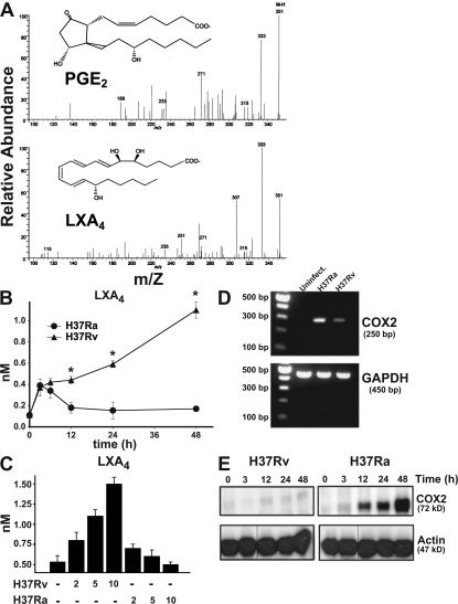
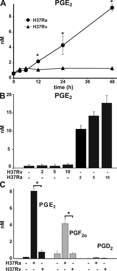
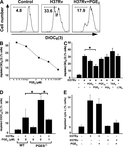
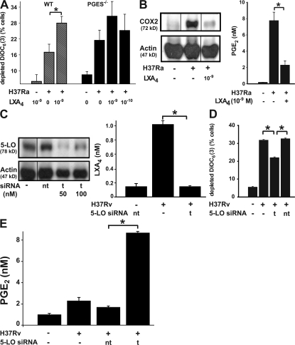
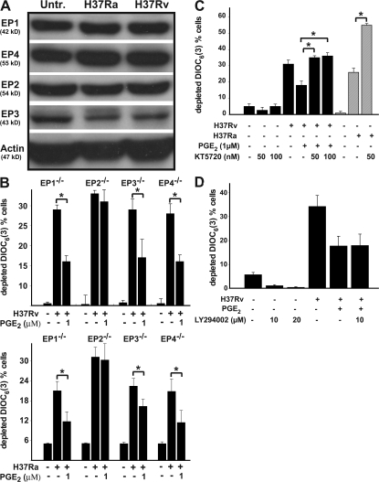
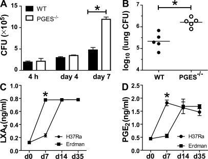
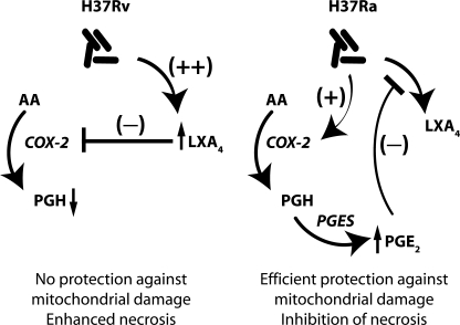
Virulent Mycobacterium tuberculosis (Mtb) induces a maladaptive cytolytic death modality, necrosis, which is advantageous for the pathogen. We report that necrosis of macrophages infected with the virulent Mtb strains H37Rv and Erdmann depends on predominant LXA(4) production that is part of the antiinflammatory and inflammation-resolving action induced by Mtb. Infection of macrophages with the avirulent H37Ra triggers production of high levels of the prostanoid PGE(2), which promotes protection against mitochondrial inner membrane perturbation and necrosis. In contrast to H37Ra infection, PGE(2) production is significantly reduced in H37Rv-infected macrophages. PGE(2) acts by engaging the PGE(2) receptor EP2, which induces cyclic AMP production and protein kinase A activation. To verify a role for PGE(2) in control of bacterial growth, we show that infection of prostaglandin E synthase (PGES)(-/-) macrophages in vitro with H37Rv resulted in significantly higher bacterial burden compared with wild-type macrophages. More importantly, PGES(-/-) mice harbor significantly higher Mtb lung burden 5 wk after low-dose aerosol infection with virulent Mtb. These in vitro and in vivo data indicate that PGE(2) plays a critical role in inhibition of Mtb replication.


Figures

Figure 1.
Figure 1.
LXA4 and COX2 production of human Mφ infected with H37Rv and H37Ra. (A) LC-MS-MS of endogenous PGE2 (top) and LXA4 (bottom) produced by H37Ra- (MOI 10) and H37Rv- (MOI 10) infected human Mφ, respectively, at 24 h. The spectrum is a representative LC-MS-MS (n = 3). The prominent ions and relative intensity matched with authentic PGE2 and LXA4 under these LC-MS-MS conditions. (B) LXA4 production in human Mφ infected with H37Rv and H37Ra (MOI 10:1) at 0–48 h. Differences in LXA4 concentrations in supernatants from H37Ra- and H37Rv-infected Mφ are statistically significant (*, P < 0.002; n = 3). (C) LXA4 accumulation at 48 h in Mφ supernatants infected with H37Ra and H37Rv (MOI 2, 5, and 10:1). The differences in LXA4 production induced by H37Ra and H37Rv are statistically significant at all MOIs (P < 0.01; n = 3). (D) COX2 mRNA accumulation in Mφ infected with H37Ra or H37Rv (MOI 10:1; uninfect, uninfected). (E) Production of COX2 protein in Mφ infected with H37Ra and H37Rv (MOI 10:1) at different time points. In all studies, n represents the number of independent experiments and the error bars represent SE. Black lines indicate that intervening lanes have been spliced out.
Figure 2.
Figure 2.
Prostanoid production of Mφ infected with Mtb. (A) PGE2 production of human Mφ 0–48 h after infection with H37Ra and H37Rv (MOI 10:1). Differences in PGE2 concentrations in supernatants from H37Ra- and H37Rv-infected Mφ are statistically significant (*, P < 0.002; n = 3). (B) PGE2 production by Mφ infected with H37Ra and H37Rv (MOI 2:1–10:1). The differences in PGE2 production induced by H37Ra in comparison to H37Rv are statistically significant at all MOIs (*, P < 0.01; n = 3). (C) Quantification of PGE2, PGF, and PGD2 in 48-h supernatants from human Mφ (5 × 10⁶ /ml) infected with H37Ra and H37Rv (MOI 10:1; *, P < 0.002; n = 4). Data are presented as means ± SE. In all studies, n represents the number of independent experiments.
Figure 3.
Figure 3.
Effect of exogenous prostanoids produced by Mtb-infected Mφ on H37Rv-induced mitochondrial cationic dye release. (A) PGE2 blocks DiOC6(3) release from mitochondria of human Mφ infected with H37Rv (MOI 5:1; 48 h). A fluorescence-activated cell sorter diagram is shown. (B) Down-regulation of cationic dye release by increasing concentrations of PGE2 (2–10 μM) is statistically significant at all PGE2 concentrations (P < 0.01; n = 6). (C) Effect of 1 μM of various prostanoids on H37Rv-induced mitochondrial DiOC6(3) release of Mφ. Only PGE2 inhibition of cationic dye release from the mitochondria is statistically significant (*, P < 0.007; n = 3). PGF has borderline activity. The concentration of LTB4 is 0.1 μM. (D) H37Ra (MOI 5:1)-induced DiOC6(3) release from mitochondria of WT and PGES−/− mouse Mφ infected in the absence and presence of 1 μM PGE2. At 48 h, cationic dye release from the mitochondria was measured (*, P < 0.006; n = 5). (E) 1 μM PGE2 does not alter H37Ra- and H37Rv-induced cytochrome c release from the mitochondria (not significant; n = 3). Data are presented as means ± SE. In all studies, n represents the number of independent experiments.
Figure 4.
Figure 4.
Effect of LXA4 on Mtb-infected Mφ. (A, left) addition of 10−9 M LXA4 to H37Ra-infected human Mφ (MOI 10:1) enhances mitochondrial cationic dye release (*, statistically significant difference; n = 3; P = 0.04). (A, right) LXA4 by itself is ineffective. Addition of 10−9 and 10−10 M LXA4 to PGES−/− mouse Mφ infected with H37Ra (MOI 5:1) does not affect mitochondrial cationic dye release (not significant; n = 3). (B, left) Production of COX2 protein by H37Ra (MOI 10:1)-infected Mφ is inhibited by the addition of 10−9 M LXA4. Western analysis of Mφ extracts. The actions of LXA4 were mimicked by its metabolic stable analogue (not depicted). (B, right) PGE2 production by Mφ (*, P < 0.001; n = 3). Black lines indicate that intervening lanes have been spliced out. (C and D) Targeted silencing (t) of the 5-LO gene (C, ∼70% inhibition at t 50) abrogates production of LXA4 by H37Rv-infected Mφ compared with nontargeted (nt) silencing (D, left, 50 nM siRNA; P = 0.001; n = 3) and reduces mitochondrial cationic dye release after H37Rv infection (D, right; nt, nontargeted; P < 0.04; n = 3). Black lines indicate that intervening lanes have been spliced out. (E) Targeted silencing of the 5-LO gene reconstitutes production of PGE2 after infection of Mφ with H37Rv, indicating that block of PGE2 production is caused by the action of LXA4 (P < 0.003; n = 3). Data are presented as means ± SE. In all studies, n represents the number of independent experiments (*, statistically significant).
Figure 5.
Figure 5.
EP2 mediates PGE2-dependent protection against cationic mitochondrial dye release. Data are presented as means ± SE. (A) EP1, EP2, EP3, and EP4 are constitutively expressed in human Mφ. Their expression is not increased by either H37Ra or H37Rv infection. (B) EP2−/− Mφ fail to respond to PGE2 by down-regulating DiCO6(3) release from the mitochondria infected with H37Rv (top) or with H37Ra (bottom), indicating that EP2 mediates the protective function of PGE2. Mφ from EP1, EP3, and EP4 −/− mice were equally responsive to 1 μM PGE2 (*, statistically significant; P < 0.01; n = 5). (C) The cAMP-dependent PKA inhibitor KT5720 abrogates inhibition of mitochondrial cationic dye release by PGE2 (black columns; P < 0.01; n = 3). Addition of KT5720 to H37Ra-infected (MOI 10:1) Mφ enhanced Mφ necrosis (gray columns; *, P < 0.01; n = 3). (D) The PI3K inhibitor LY294002 does not abrogate inhibition of mitochondrial cationic dye release by PGE2 (not significant; n = 3). In all studies, n represents the number of independent experiments.
Figure 6.
Figure 6.
Mycobacterial burden of PGES−/− and WT Mφ in vitro and in vivo. (A) Mycobacterial burden after 4 h (inoculum), 4 d, and 7 d of PGES−/− and WT Mφ in vitro infected with H37Rv (MOI 10:1). The difference in the bacterial burden was significant at 4 and 7 d after infection (*, P < 0.03). (B) 5 wk after aerosol infection, CFUs were determined by plating of homogenized lung tissue as indicated on the ordinate. The difference in mycobacterial burden in the lungs of PGES−/− versus WT mice is statistically significant (*, P = 0.002; five mice per group). (C and D) Induction of LXA4 and PGE2 production during the course of mycobacterial infection. WT mice were infected by aerosol exposure with virulent (Erdmann) or avirulent (H37Ra). LXA4 (C) and PGE2 (D) measured by ELISA in the sera at 7, 14, and 35 d after infection (*, P < 0.01; three mice per time points). These results are representative of two independent experiments. The error bars represent SE.
Figure 7.
Figure 7.
Infection of Mφ with the virulent H37Rv predominantly induces LXA4 production and block of COX2 and PGE2 production, which might lead to necrotic cell lysis and spread of the infection. In contrast, Mφ infected with avirulent H37Ra produce larger amounts of PGE2 which blocks LXA4 production and supports Mφ apoptosis and containment of the Mtb. Predominant production of either PGE2 or LXA4 by Mφ infected with H37Ra or H37Rv, respectively, is supported by the fact that PGE2 inhibits LXA4 production and vice versa.

References

    1. Corbett, E.L., C.J. Watt, N. Walker, B.G. Williams, M.C. Raviglione, and C. Dye. 2003. The growing burden of tuberculosis: global trends, and interactions with the HIV epidemic. Arch. Intern. Med. 163:1009–1021. - PubMed
    1. Dannenberg, A.M., and G.A. Rook. 1994. Pathogenesis of pulmonary tuberculosis: an interplay of tissue-damaging and macrophage-activating immune responses – dual mechanisms that control bacillary multiplication. In Tuberculosis. Pathogenesis, Protection and Control. Bloom B R, editor. American Society for Microbiology, Washington DC. 459–484.
    1. Leemans, J.C., N.P. Juffermans, S. Florquin, N. van Rooijen, M.J. Verwordeldonk, A. Verbon, S.J.H. van Deventer, T. van der Poll. 2001. Depletion of alveolar macrophages exerts protective effects in pulmonary tuberculosis in mice. J. Immunol. 166:4604–4611. - PubMed
    1. Russell, D.G., H.C. Mwandumba, and E.E. Rhoades. 2002. Mycobacterium and the coat of many lipids. J. Cell Biol. 158:421–426. - PMC - PubMed
    1. Sturgill-Koszycki, S., U.E. Schaible, and D.G. Russell. 1996. Mycobacterium-containing phagosomes are accessible to early endosomes and reflect a transitional state in normal phagosome biogenesis. EMBO J. 15:6960–6968. - PMC - PubMed

Publication types

MeSH terms