

Calcium and apoptosis: ER-mitochondria Ca2+ transfer in the control of apoptosis
- PMID: 18955969
- PMCID: PMC2844952
- DOI: 10.1038/onc.2008.308
Calcium and apoptosis: ER-mitochondria Ca2+ transfer in the control of apoptosis
Abstract
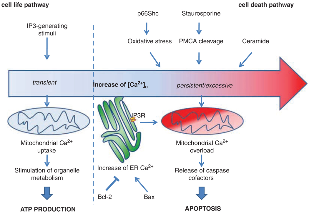
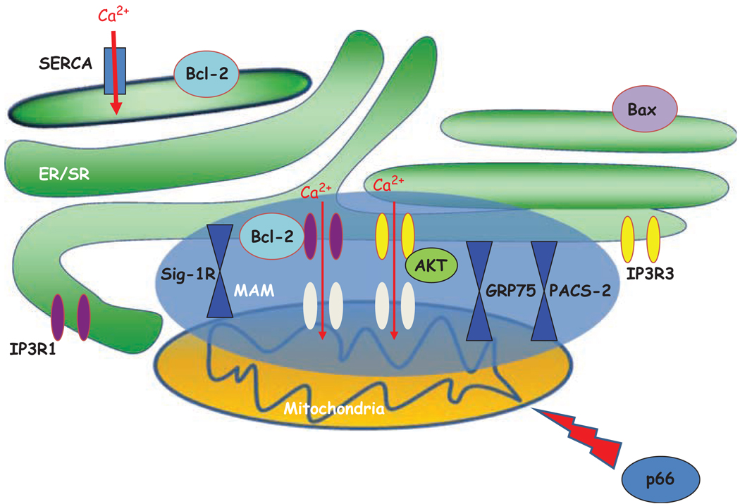
There is a growing consensus that the various forms of cell death (necrosis, apoptosis and autophagy) are not separated by strict boundaries, but rather share molecular effectors and signaling routes. Among the latter, a clear role is played by calcium (Ca(2+)), the ubiquitous second messenger involved in the control of a broad variety of physiological events. Fine tuning of intracellular Ca(2+) homeostasis by anti- and proapoptotic proteins shapes the Ca(2+) signal to which mitochondria and other cellular effectors are exposed, and hence the efficiency of various cell death inducers. Here, we will review: (i) the evidence linking calcium homeostasis to the regulation of apoptotic, and more recently autophagic cell death, (ii) the discussion of mitochondria as a critical, although not unique checkpoint and (iii) the molecular and functional elucidation of ER/mitochondria contacts, corresponding to the mitochondria-associated membrane (MAM) subfraction and proposed to be a specialized signaling microdomain.
Figures
References
-
- Alnemri ES, Livingston DJ, Nicholson DW, Salvesen G, Thornberry NA, Wong WW, et al. Human ICE/CED-3 protease nomenclature. Cell. 1996;87:171. - PubMed
-
- Assefa Z, Bultynck G, Szlufcik K, Nadif KN, Vermassen E, Goris J, et al. Caspase-3-induced truncation of type 1 inositol trisphosphate receptor accelerates apoptotic cell death and induces inositol trisphosphate-independent calcium release during apoptosis. J Biol Chem. 2004;279:43227–43236. - PubMed
-
- Baehrecke EH. Autophagy: dual roles in life and death? Nat Rev Mol Cell Biol. 2005;6:505–510. - PubMed
-
- Baffy G, Miyashita T, Williamson JR, Reed JC. Apoptosis induced by withdrawal of interleukin-3 (IL-3) from an IL-3-dependent hematopoietic cell line is associated with repartitioning of intracellular calcium and is blocked by enforced Bcl-2 oncoprotein production. J Biol Chem. 1993;268:6511–6519. - PubMed
-
- Baines CP, Kaiser RA, Purcell NH, Blair NS, Osinska H, Hambleton MA, et al. Loss of cyclophilin D reveals a critical role for mitochondrial permeability transition in cell death. Nature. 2005;434:658–662. - PubMed
Publication types
MeSH terms
Substances
Grants and funding
LinkOut - more resources
Full Text Sources
Other Literature Sources
Miscellaneous
